Synovial Joints
830 likes | 996 Vues
Synovial Joints. PTP 521 Musculoskeletal Diseases and Dysfunctions. Objectives. Review the embryological development of joints Review the components of a joint Discuss the histological makeup of joint components Discuss diseases and disorders that stem from particular joint dysfunctions

Synovial Joints
E N D
Presentation Transcript
Synovial Joints PTP 521 Musculoskeletal Diseases and Dysfunctions
Objectives • Review the embryological development of joints • Review the components of a joint • Discuss the histological makeup of joint components • Discuss diseases and disorders that stem from particular joint dysfunctions • Discuss healing rates of joint components
Embryonic Development of Human Joints • Joints appear as intervals of less concentrated mesenchymal cells • These cells become flattened in the center. • At the ends, the flattened cells are continuous with the investing perichondrium • The perichondrial investment becomes the joint capsule • 6th week: joints develop • 8th week: joints resemble adult joints
As soon as joint cavity appears during development, it contains watery fluid • Joint capsule develops into an outer fibrous portion lined with an inner portion which is the highly vascularized synovial membrane • Articular Cartilage is formed from the original cartilagenous template
Articular Cartilage Embryological Development: • Develops during week 5 • Mesenchymal cells condense, enlarge, proliferate to form compact mass • Cells (Chondrocytes) become rounded, secrete matrix
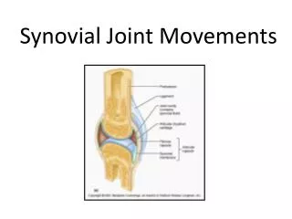
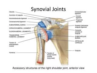
Synovial Joint Components • Joint Capsule • Synovium • Synovial Fluid • Articular Cartilage
Joint Capsule • Completely encloses the synovial joint and covers all surfaces except for articulating surfaces and contact surface of any intra-articular structure such as a meniscus • Hydrated, water content of ~70% • Forms a cuff around the joint with collagen fibers, Type I • Variability in thickness and attachment of capsule, some are thin attachments and some are thick • Capsular redundancy D. Fiber diameter varies, organized in parallel bundles • Capsular thickenings or intrinsic ligaments are present, reinforce the capsule.
Capsule Inflammatory Response • High potential for adhesion formation • Loss of Glycoaminoglycogen content and water • Fiber-fiber distance decreases, folds of synovial fluid adhere together limiting mobility
Capsule Injury • Joint Effusion: Signs: joint moves to position of comfort Symptoms: tight sensation in joint B.Capsular Fibrosis: Thickened capsule, collagen fibers have decreased extensibility Signs: decrease in ROM, capsular pattern present
Joint Mechanoreceptors Sensory receptor located within the joint capsule and articular ligaments Responds to mechanical pressure or deformation • 4 Main types • PacinianCorpusles • RuffiniCorpusles • Golgi – like tendon organs • Free nerve endings – type C
Joint Mechanoreceptors:Capsule and Ligaments • RuffiniCorpuscles • Located superficial joint capsules • Postural receptors • Low threshold, easy to stimulate, slow to adapt • Stimulated by a change in mechanical stress about the joint • Give information on static and dynamic position, sense of velocity, direction and amplitude of movement • Responds by increasing tone in muscle being stretched, relaxing antagonistic muscle • Pacinian Corpuscles • Located in small groups in joint capsule • Low threshold, rapidly adapting, active with oscillation techniques rather than traction • Stimulated by sudden changes in movement, pressure changes within the joint • Give a sense of joint acceleration and deceleration • Increases tone in muscle being stretched and relaxes antagonistic muscle
Joint Mechanoreceptors • Golgi-Tendon Like • Inhibitive • NOT in joint capsule but rather found in ligaments • High threshold, slowly adapting, more active with high velocity techniques • Stimulated by stretch at end range • Function is to prevent excessive stresses at joint by reflexively inhibiting muscle • Sense of dynamic movement and direction of movement • Nociceptors: Unmyelinated free nerve endings: Type C • Found in capsule, ligament, articular fat pads, periosteum, walls of blood vessels • Burning, aching pain that is difficult to localize • tonic receptors – responds by producing a tonic muscle contraction • no significant peripheral adaptation, but central adaptation can occur(perception of pain may decrease) • High threshold, non adaptive • Stimulated by excessive movement
Joint Mechanoreceptors:Capsule and Ligaments • Pacinian Corpuscles • Located in small groups in joint capsule • Low threshold, reapidly adapting active with ocillation techniques rather than traction • Stimulated by • Ruffini • Located superficial to joint capsule • Postural receptors • Low threshold, easy to stimulate, slow to adapt • Stimulated by a change in mechanical stress about the joint • Give info on static and dynamic position, sense of velocity, direction and amplitude movement • Responds to stretch by increasing muscle tone, relaxing antagonistic mucles
Joint Mechanoreceptors • Golgi-Tendon Like • Inhibitive, Not in joint capsule but rather found in ligaments. • Stimulated by stretch at end range. • Sense of dynamic movemnent and direction of movemnet • Nociceptors: Unmyelinated free nerve endings: Type C • Found in capsule, ligament, fat pads, periosteum, walls of blood vessels • Burning aching pain that is difficult to localize • Tonic receptors-responds b producing a tonic muscle contraction • High threshold, non adaptive • Stimulated by excessive movements
Synovium Synoviocytes: Specialized layer of connective tissue cells • Covers the inner surface of the joint capsule to the margins of the articular cartilage. • Does not cover weight bearing surfaces.
Synovium:Two Layers of Cells 1) Intima • Layer of specialized fibroblasts, 1-3 deep, set within a matrix • Contain both macrophages derived type A cells and fribroblast-derived type B cells • Synthesizes hyaluronic acid and other cells which help lubricate the joint • Line the joint cavity
Subsynovial tissue • Produces collagen matrix • Fibrous, areolar, and adipose tissue • Contains lymphatics and blood vessels
Vascularity • Varies between joints • Varies within a joint • Lowest in fibrous subsynovial tissues • Greatest in areolar or adipose subsynovial tissue
Functions of the Synovium • Joint Lubrication: provides a low friction lining 2) Transportation – nutrition • Joint Stability • Other: • Regulation of intra-articular temperature • May have an anti-microbial effect
Synovium: Reaction to Injury 1) Proliferation of Surface Cells 2) Increase in vascularization 3) Gradual Fibrosis of Subsynovial Tissue 4) Granular Surface appears 5) Alteration in synovial fluid
Disease Processes 1) Post Traumatic Synovitis Pathophysiology: • Intima layers increase to 8-10 and as much as 15 • Increase in lymphatics, protein synthesis Clinical Manifestations • Signs: joint effusion • Symptoms: tight sensation around joint
Hemarthrosis • Fracture must be ruled out first • Signs: rapid swelling within 15 min to 2 hours after injury, loss of range due to increase in swelling • Symptoms: increase in pain, tight sensation
Pigmented VillonodularSynovitis • Nodules formed in the synovial tissue due to blood clots attaching to the synovium. • 1.8 cases/million • Women > Men • 20-50 years of age • Theory: auto immune response or benign neoplastic process
PVS: • During joint motion, the nodules are crushed and cause further hemorrhage. This leads to more nodules and more pain occurring. • Inflammatory signs and symptoms • Signs: Pain with compression of joint surfaces • Medical RX: synovectomy Illustrated by Dave Klemm
Rheumatoid Arthritis: Definition: chronic systemic inflammatory disease which can result in severe deformities and disabilities. http://www.emedicine.com/RADIO/topic877.htm
R.A. Cont. • Cervical spine and PIP joints of the hand are commonly involved. • Incidence: affects all races and ages. 1-2% of the adult population have RA
Risk Factors: • Women are 2-3 times more likely to develop RA than men • Age: 30 to 40 years of age is peak onset time c. RA incidence is lower in women who have had children and in women who have taken oral contraceptives
R.A. Cont. 1. Etiology: unknown 2. Pathology: 80% have a positive rheumatoid factor
R.A. Cont. 3. SX: joint stiffness 98% are stiffer after inactivity and better with activity. • Joint pain: severity of synovitis – may not be a factor at rest • Fatigue onset about 4 ½ hours after rising • Weakness out of proportion to activity
Signs: • Swelling in the small joints and tendon sheaths in the hands wrists and forefeet is most common. Can affect any area of the body. • Palmar erythema is very common, (palms and thenar eminences)
R.A. Cont. • Cool moist skin • Atrophy of muscles in hands and feet • Can get extensive joint contractures at later stages http://www.emedicine.com/RADIO/topic877.htm
Treatment goals: • Reduce pain, maintain mobility, minimize stiffness, edema and joint destruction, aggressive treatment is contraindicated.
R.A. Cont. Medical management: diagnosis is made with lab tests and is also based on history, physical examination. • Surgery is indicated if conservative care is not sufficient to control pain and maintain functional status.
American Rheumatism Association 4/7 conditions with the first 4 present at least 6 weeks 1. morning stiffness greater than 1 hour 2. arthritis in 3 or more joints with swelling 3. arthritis of hands, joints with swelling 4. symmetric arthritis 5. rheumatoid nodules 6. x-ray findings are typical for RA 7. + lab tests – serum rheumatoid factor
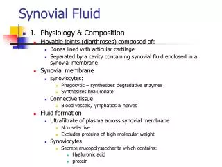
Synovial Fluid Blood plasma with additions of proteins and hyaluronic acid Analysis of synovial fluid involves process of arthrocentesis which is the surgical withdrawal of fluid from the cavity
Categories 1) Normal 2) Group I: Non-inflammatory 3) Group II: Inflammatory 4) Group III: Purulent 5) Group IV: Hemorrhagic
Group IV: Hemorrhagic: presence of blood in the synovial fluid. If blood is present at all, it is in this grouping. • Examples: trauma, fracture, charcot joint, hemophilia, tumor, joint prosthesis, sickle cell trait or disease, internal derangement, major ligament tear • Other: Fat – indicates a fracture • Cloudy Synovial Fluid: usually caused by leukocytes. If greater than 2000, will be concerned about an inflammatory joint disease
Articular Cartilage: • Composition: Water, Collagen, Proteoglycans • Nutritional Supply: occurs with diffusion and osmosis • Functions: Carry a heavy load
Histological Composition • Cells: • Chondroblasts: young cell • Fibroblasts • Fibrocytes • Chondrocytes 2-5% of volume,
Chondrocyte Function Synthesizes Matrix composed of proteoglycans, growth factors, cytokines, collagen fibers Anabolic activity and catabolic activity Common for cells to be multinucleated Every cell contains a monocillium (mechanotransductory function)
Articular Cartilage Zones • Zone 1: superficial tangential zone • Zone 2: transitional zone • Zone 3: deep radial layer • Zone 4: cartilage zone • tidemark
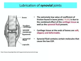
Joint Lubrication Boundary Lubrication: • low load, • prevents direct surface contact • eliminates surface wear, • dependent on Lubricin
Fluid Film Lubrication: high load A. squeeze film lubrication: perpendicular movement, high loads, short duration occurs with synovial fluid viscosity Zachazewski JE, Magee DJ, Quillen WS. Athletic Injuries and Rehabilitation. W.B. Saunders Co. 1996
Fluid Film Lubrication B. hydrodynamic lubrication: tangential movement, lifting pressure Zachazewski et al, 1996
Fluid Film Lubrication C. Elastohydrodynamic: mixed mode of lubrication • Articular cartilage operates under this type of lubrication Zachazewski et al, 1996
Fluid Film Lubrication D. Boosted Lubrication: mixture of both boundary and squeeze film lubrication Zachazewski et al, 1996