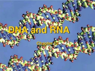
DNA and RNA
1.96k likes | 2.15k Vues
DNA and RNA. DNA. How do genes work? What are they made of, and how do they determine the characteristics of organisms? Are genes single molecules, or are they longer structures made up of many molecules?

DNA and RNA
E N D
Presentation Transcript
DNA • How do genes work? • What are they made of, and how do they determine the characteristics of organisms? • Are genes single molecules, or are they longer structures made up of many molecules? • In the middle of the 1900s, questions like these were on the minds of biologists everywhere
DNA • To truly understand genetics, biologists first had to discover the chemical nature of the gene • If the structures that carry genetic information could be identified, it might be possible to understand how genes control the inherited characteristics of living things
Griffith and Transformation • Like many stories in science, the discovery of the molecular nature of the gene began with an investigator who was actually looking for something else • In 1928, British scientist Frederick Griffith was trying to figure out how bacteria make people sick • More specifically, Griffith wanted to learn how certain types of bacteria produce a serious lung disease known as pneumonia
Griffith and Transformation • Griffith had isolated two slightly different strains, or types, of pneumonia bacteria from mice • Both strains grew very well in culture plates in his lab, but only one of the strains caused pneumonia • The disease-causing strain of bacteria grew into smooth colonies on culture plates, whereas the harmless strain produced colonies with rough edges • The differences in appearance made the two strains easy to distinguish
Griffith's Experiments • When Griffith injected mice with the disease-causing strain of bacteria, the mice developed pneumonia and died • When mice were injected with the harmless strain, they didn't get sick at all • Griffith wondered if the disease-causing bacteria might produce a poison
Griffith's Experiments • To find out, he took a culture of these cells, heated the bacteria to kill them, and injected the heat-killed bacteria into mice • The mice survived, suggesting that the cause of pneumonia was not a chemical poison released by the disease-causing bacteria
Griffith's ExperimentsTransformation • Griffith injected mice with four different samples of bacteria • When injected separately, neither heat-killed, disease-causing bacteria nor live, harmless bacteria killed the mice • The two types injected together, however, caused fatal pneumonia • From this experiment, biologists inferred that genetic information could be transferred from one bacterium to another
Transformation • Griffith's next experiment produced an amazing result • He mixed his heat-killed, disease-causing bacteria with live, harmless ones and injected the mixture into mice • By themselves, neither should have made the mice sick • But to Griffith's amazement, the mice developed pneumonia and many died • When he examined the lungs of the mice, he found them filled not with the harmless bacteria, but with the disease-causing bacteria • Somehow the heat-killed bacteria had passed their disease-causing ability to the harmless strain • Griffith called this processtransformationbecause one strain of bacteria (the harmless strain) had apparently been changed permanently into another (the disease-causing strain)
Transformation • Griffith hypothesized that when the live, harmless bacteria and the heat-killed bacteria were mixed, some factor was transferred from the heat-killed cells into the live cells • That factor, he hypothesized, must contain information that could change harmless bacteria into disease-causing ones • Furthermore, since the ability to cause disease was inherited by the transformed bacteria's offspring, the transforming factor might be a gene
Avery and DNA • In 1944, a group of scientists led by Canadian biologist Oswald Avery at the Rockefeller Institute in New York decided to repeat Griffith's work • They did so to determine which molecule in the heat-killed bacteria was most important for transformation • If transformation required just one particular molecule, that might well be the molecule of the gene
Avery and DNA • Avery and his colleagues made an extract, or juice, from the heat-killed bacteria • They then carefully treated the extract with enzymes that destroyed proteins, lipids, carbohydrates, and other molecules, including the nucleic acid RNA • Transformation still occurred • Obviously, since these molecules had been destroyed, they were not responsible for the transformation
Avery and DNA • Avery and the other scientists repeated the experiment, this time using enzymes that would break down DNA • When they destroyed the nucleic acid DNA in the extract, transformation did not occur • There was just one possible conclusion • DNA was the transforming factor • Avery and other scientists discovered that the nucleic acid DNA stores and transmits the genetic information from one generation of an organism to the next
The Hershey-Chase Experiment • Scientists are a skeptical group • It usually takes several experiments to convince them of something as important as the chemical nature of the gene • The most important of these experiments was performed in 1952 by two American scientists, Alfred Hershey and Martha Chase • They collaborated in studying viruses, nonliving particles smaller than a cell that can infect living organisms
Bacteriophages • One kind of virus that infects bacteria is known as a bacteriophage, which means “bacteria eater” • Bacteriophages are composed of a DNA or RNA core and a protein coat • When a bacteriophage enters a bacterium, the virus attaches to the surface of the cell and injects its genetic information into it • The viral genes act to produce many new bacteriophages, and they gradually destroy the bacterium • When the cell splits open, hundreds of new viruses burst out
Radioactive Markers • Hershey and Chase reasoned that if they could determine which part of the virus—the protein coat or the DNA core—entered the infected cell, they would learn whether genes were made of protein or DNA • To do this, they grew viruses in cultures containing radioactive isotopes of phosphorus-32 (32P) and sulfur-35 (35S) • This was a clever strategy because proteins contain almost no phosphorus and DNA contains no sulfur • The radioactive substances could be used as markers • If 35S was found in the bacteria, it would mean that the viruses' protein had been injected into the bacteria • If 32P was found in the bacteria, then it was the DNA that had been injected
Radioactive Markers • The two scientists mixed the marked viruses with bacteria • Then, they waited a few minutes for the viruses to inject their genetic material • Next, they separated the viruses from the bacteria and tested the bacteria for radioactivity • Nearly all the radioactivity in the bacteria was from phosphorus (32P), the marker found in DNA • Hershey and Chase concluded that the genetic material of the bacteriophage was DNA, not protein
NUCLEIC ACIDS • Types: • DNA: Deoxyribonucleic Acid • RNA: Ribonucleic Acid • mRNA • rRNA • tRNA
DNA • Two primary functions: • Stores and uses information to direct the activities of the cell • Copy itself exactly for new cells that are created • Controls the production of proteins within the cell • These proteins form the structural units of cells and control all chemical processes (enzymes) within cells • We have inherited DNA from our biological parents and we will pass our DNA to our biological offspring
The Components and Structure of DNA • You might think that knowing genes were made of DNA would have satisfied scientists, but that was not the case at all • Instead, they wondered how DNA, or any molecule for that matter, could do the three critical things that genes were known to do: • First,genes had to carry information from one generation to the next • Second, they had to put that information to work by determining the heritable characteristics of organisms • Third, genes had to be easily copied, because all of a cell's genetic information is replicated every time a cell divides • For DNA to do all of that, it would have to be a very special molecule indeed
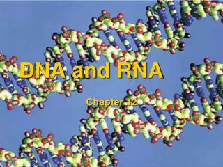
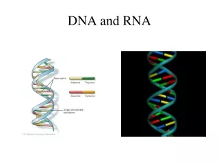
The Components and Structure of DNA • DNA is a long molecule made up of units called nucleotides • As the figure below shows, each nucleotide is made up of three basic components: • 5-carbon sugar called deoxyribose • Phosphate group • Nitrogenous (nitrogen-containing) base • There are four kinds of nitrogenous bases in DNA: • Two of the nitrogenous bases, adenine and guanine, belong to a group of compounds known as purines • The remaining two bases, cytosine and thymine, are known as pyrimidines • Purines have two rings in their structures, whereas pyrimidines have one ring
STRUCTURE OF DNA • DNA is a polymer that is composed of repeating subunits (monomers) called nucleotides • DNA molecule consists of two long strands, each of which is a chain of nucleotide monomers • Nucleotide: has three parts • Deoxyribose: a five-carbon sugar molecule • A phosphate group • A nitrogen base: can have one of four types • Purine types: adenine or guanine • Pyrimidine types: thymine or cytosine
The Components and Structure of DNA • DNA Nucleotides: DNA is made up of a series of monomers called nucleotides • Each nucleotide has three parts: • Deoxyribose molecule • Phosphate group • Nitrogenous base: • There are four different bases in DNA: adenine, guanine, cytosine, and thymine.
The Components and Structure of DNA • The backbone of a DNA chain is formed by sugar and phosphate groups of each nucleotide • The nitrogenous bases stick out sideways from the chain • The nucleotides can be joined together in any order, meaning that any sequence of bases is possible
The Components and Structure of DNA • In the 1940s and early 1950s, the leading biologists in the world thought of DNA as little more than a string of nucleotides • The four different nucleotides, like the 26 letters of the alphabet, could be strung together in many different ways, so it was possible they could carry coded genetic information • However, so could many other molecules, at least in principle • Was there something more to the structure of DNA?
The Components and Structure of DNAChargaff's Rules • One of the puzzling facts about DNA was a curious relationship between its nucleotides • Years earlier, Erwin Chargaff, an American biochemist, had discovered that the percentages of guanine [G] and cytosine [C] bases are almost equal in any sample of DNA • The same thing is true for the other two nucleotides, adenine [A] and thymine [T], as shown in the table • The observation that [A] = [T] and [G] = [C] became known as Chargaff's rules • Despite the fact that DNA samples from organisms as different as bacteria and humans obeyed this rule, neither Chargaff nor anyone else had the faintest idea why
The Components and Structure of DNAChargaff's Rules • Chargaff's Rules: • Erwin Chargaff showed that the percentages of guanine and cytosine in DNA are almost equal • The same is true for adenine and thymine.
The Components and Structure of DNA X-Ray Evidence • In the early 1950s, a British scientist named Rosalind Franklin began to study DNA • She used a technique called X-ray diffraction to get information about the structure of the DNA molecule • Aiming a powerful X-ray beam at concentrated DNA samples, she recorded the scattering pattern of the X-rays on film • Franklin worked hard to make better and better patterns from DNA until the patterns became clear
The Components and Structure of DNA X-Ray Evidence • By itself, Franklin's X-ray pattern does not reveal the structure of DNA, but it does carry some very important clues • The X-shaped pattern in the photograph in the image below shows that the strands in DNA are twisted around each other like the coils of a spring, a shape known as a helix • The angle of the X suggests that there are two strands in the structure • Other clues suggest that the nitrogenous bases are near the center of the molecule
The Components and Structure of DNA X-Ray Evidence • X-Ray Diffraction Image of DNA : • X-ray diffraction is the method that Rosalind Franklin used to study DNA.
The Double Helix • The same time that Franklin was continuing her research, Francis Crick, a British physicist, and James Watson, an American biologist, were trying to understand the structure of DNA by building three-dimensional models of the molecule • Their models were made of cardboard and wire • They twisted and stretched the models in various ways, but their best efforts did nothing to explain DNA's properties
The Double Helix • Then, early in 1953, Watson was shown a copy of Franklin's remarkable X-ray pattern • The effect was immediate • In his book The Double Helix, Watson wrote: “The instant I saw the picture my mouth fell open and my pulse began to race” • Using clues from Franklin's pattern, within weeks Watson and Crick had built a structural model that explained the puzzle of how DNA could carry information, and how it could be copied • They published their results in a historic one-page paper in April of 1953 • Watson and Crick's model of DNA was a double helix, in which two strands were wound around each other
STRUCTURE OF DNA • Double helix: • Each nucleotide (deoxyribose, phosphate, and a nitrogen base) bonds (sugar to phosphate) to other nucleotides to form a long strand • Nitrogen bases not involved in this bonding • Two of these strands bonded together (H bonds between the nitrogen bases) form a molecule of DNA • Hydrogen bond: type of chemical bond in which atoms share a hydrogen nucleus (one proton) • Between purine and pyrimidine • Sugar and phosphate not involved in this bonding • The two strands twist around a central axis to form a spiral structure called a double helix (twisted ladder) • Sides of the ladder are formed by alternating sugar and phosphate units • Rungs of the ladder consist of bonded pairs (H bonds) of nitrogen bases • Rungs are of uniform length because a purine bonds with a pyrimidine • Adenine (A) always bonds with Thymine (T): 2 H bonds • Cytosine (C) always bonds with Guanine (G): 3 H bonds • Right hand twist with each turn consisting of 10 base pairs
STRUCTURE OF DNA • The sequential arrangement of nitrogen bases along one strand is the exact complement of the sequential arrangement of bases on the adjacent strand • Example of complementary strands: • A-T • C-G • T-A • G-C
The Double Helix • A double helix looks like a twisted ladder or a spiral staircase • When Watson and Crick evaluated their DNA model, they realized that the double helix accounted for many of the features in Franklin's X-ray patternbut did not explain what forces held the two strands together • They then discovered that hydrogen bonds could form between certain nitrogenous bases and provide just enough force to hold the two strands together
The Double Helix • Hydrogen bonds can form only between certain base pairs—adenine and thymine, and guanine and cytosine • Once they saw this, they realized that this principle, called base pairing, explained Chargaff's rules • Now there was a reason that [A] = [T] and [G] = [C] • For every adenine in a double-stranded DNA molecule, there had to be exactly one thymine molecule • For each cytosine molecule, there was one guanine molecule
The Double Helix • DNA Structure: • DNA is a double helix in which two strands are wound around each other • Each strand is made up of a chain of nucleotides • The two strands are held together by hydrogen bondsbetween adenine and thymine and between guanine and cytosine
Chromosomes and DNA Replication • DNA is present in such large amounts in many tissues that it's easy to extract and analyze • But where is DNA found in the cell? • How is it organized? • Where are the genes that Mendel first described a century and a half ago?
DNA and Chromosomes • Prokaryotic cells lack nuclei and many of the organelles found in eukaryotes • Their DNA molecules are located in the cytoplasm • Most prokaryotes have a single circular DNA molecule that contains nearly all of the cell's genetic information • This large DNA molecule is usually referred to as the cell's chromosome