OSSEOUS TISSUE
OSSEOUS TISSUE. SKELETAL STRUCTURE. Skeletal System. 206 bones, cartilage, ligaments, and connective tissues Functions: support provides a rigid framework storage calcium & phosphorus lipids production of blood cells formed in red marrow protection brain is encased in skull

OSSEOUS TISSUE
E N D
Presentation Transcript
OSSEOUS TISSUE SKELETAL STRUCTURE
Skeletal System • 206 bones, cartilage, ligaments, and connective tissues • Functions: • support • provides arigid framework • storage • calcium & phosphorus • lipids • production of blood cells • formed in red marrow • protection • brain is encased in skull • heart and lungs are surrounded by boney sternum and rib cage • leverage • allows for movement due to interaction of muscular & skeletal systems • acid-base balance • absorbs or releases alkaline salts
Divisions of the Skeletal System • Axial skeleton • consists of bones forming axis of the body • Skull • Hyoid • Sternum • Ribs • Vertebrae • sacrum & cocyx • auditory ossicles (not a part of either; but put here by convention) • Appendicularskeleton • consists of bones that anchor appendages to axial skeleton • upper & lower extremities, shoulder and pelvic girdles
Types of Bones • Long • Flat • Short • Irregular • Sesamoid • Sutural
Long Bones • longer than wide • function as levers • act on skeletal muscles to produce movements • found in appendages • fingers & toes
Short Bones • boxy & small • nearly cube-shape • found in wrist-carpals • ankle-tarsals • limited movements
Flat Bones • thin • roughly parallel surfaces • found in the roof of the skull • sternum, ribs & scapula • enclose & protect soft organs • provide broad surfaces for muscle attachment
Irregular Bones • bones that do not fall into any other category • varied, complex shapes, sizes & surface features • vertebrae, sacrum, coccyx, temporal, sphenoid, ethmoid, zygomatic, maxilla, mandible, palatine, inferior nasal concha, & hyoid
Sesamoid Bones • shaped like sesame seeds develop in areas where there is a great deal of friction • most only a few mms number in each person differs • patella present in everyone
Sutural Bones • also called Wormian bones • small • located in sutures • classified by location-not by shape
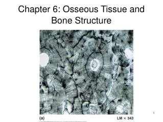
Bone Composition • Osseous Tissue • supporting connective tissue • Composed of an extra cellular matrix and specialized cells • give flexibility • two types • compact ordense bone • dense, hard, & relatively solid • forms protective exterior of all bones • spongy or cancellous bone • found inside most compact bone • very porous • full of tiny holes forming open networks of struts & plates • lighter than compact bone • reduces skeletal weight • makes it easier for muscles to move bones
Extracellular Matrix • composed of collagen fibers & ground substance • hardened by inorganic calcium phosphate deposits • called mineralization or calcification • solid calcium phosphate salts deposited around protein fibers • Calcium phosphate makes up 2/3rd of bone weight • Calcium phosphate + calcium hydroxide hydroxyapatite-Ca10(PO4)6(OH)2 • Calcium phosphate is hard, brittle & inflexible • can withstand compression • Collagen fibers are stronger than steel, flexible- can be twisted & bent • not good at being compressed • collagen makes a frame around which calcium minerals deposit • combination makes bone flexible, strong & resistant to shattering
Bone Cell Types • Osteogenic cells • stem cells produce other bone cells • found in cellular layer of periosteum, endosteum & central canals • continually divide • only bone cell that can divide • Osteoblasts • bone-forming cells • make organic matter of bone matrix • Osteocytes • mature bone cells • most of bone cell population • former osteoblasts that have become trapped in matrix they have deposited • Osteoclasts • bone destroying cells
Osteocytes • cannot divide • function to maintain & monitor protein & mineral content of matrix • participate in bone repair by converting back into osteoblasts or osteogeneic cells at the site of injury • sense strain & regulate bone remodeling
Osteoclasts • bone dissolving cells • function to remove bone by osteolysis • secrete acids & proteolytic enzymes which degrade minerals & fibers and dissolve boney matrix • releases matrix components into the blood restoring calcium and phosphorus concentrations in body fluids
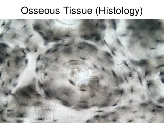
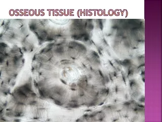
Types of Bone Tissue • Compact Bone • dense • covers exterior of all bones • Spongy Bone • cancellous • trabecular • inside compact bone • lighter
Compact Bone • basic functional unit -osteon or Haversian system. • osteocytes are arranged in concentric circles or layers-lamellae • around a central or Haversian canal • runs parallel to surface • contains blood vessels • perforating central canal are Volkmann’s canals • run perpendicular to surface • canaliculi run through layers • connect osteocytes to each other • interstitial lamellae fill spaces between
Spongy Bone • matrix composition-same • osteocytes, canalicui & lamellae-different arrangements • has no osteons • matrix forms plates or struts called trabeculae (little beams) • form a thin, branching open network filled with red bone marrow • makes bone lighter
Bone Type & Bone Tissue Type Location • the relationship between compact & spongy bone and the relative proportions of each varies with bone shape & with the function of the bone
Long Bone Structure • Diaphysis or shaft-long & cylindrical • Outside made of dense bone • medullary canal or marrow cavity isfilled with marrow • Yellow bone marrow isdominated by fat cells & red marrow is responsible for forming blood cells • Epiphysis-expanded extremities at either end of the bone • articulates with other bones-forming joints • have broad surfaces for muscle attachment. • filled with cancellous tissue surrounded by thin layer of compact bone • Metaphysis • connects diaphysis to epiphysis
Flat Bone Composition • function • provideprotection for underlying structures • broad surfaces for muscle attachment • function can be seen by structure • resembles a spongy bone sandwich • composed of 2 thin layers of compact bone covering a layer of spongy bone • bone marrow is present • there is no marrow cavity
Periosteum & Endosteum • Periosteum • covers all portions of compact bone except at joint cavities • has fibrous outer layer & an inner cellular layer • isolates bones from surrounding tissues • provides route for blood vessels & nerves • participates in bone growth & repair • continuous with other connective tissues that mesh with-tendons & ligaments • perforating or Sharpey’s fibers bond tendons & ligaments into the general structure of bone • endosteum • consists of an incomplete cellular layer • lines marrow cavities • covers trabeculae of spongy bones • lines inner surfaces of central canals • active during bone growth, repair, and remodeling
Blood & Nerve Supply • bone tissue is highly vascular • Vessels pass into the bone through the periosteum • Periosteal arteries enter via perforating canals • nutrient artery & vein • enter through a nutrient foramen located in middle of the bone
Bone Growth • new bone matrix is made through osteogenesis or ossification • process makes & releases proteins & other organic components of matrix • substance is osteoid • bone matrixbefore calcium salts have been added • calcium salts are laid down in a process called calcification
Bone Development & Growth • skeleton begins to form at 6 weeks post fertilization • does not stop until around age 25 • develops by two methods • intramembranous ossification • endochondral ossification
Intramembranous Ossification • bone forms frommesenchymeorfibrous connective tissue • produces flat bones of skull, most of the facial bones, mandible & medial part of the clavicle • bone develop within a fibrous sheet similar to dermis of the skin • bones are called dermal bones
Intramembranous Ossification Steps • Step1: Development of Ossification Center • Step 2: Calcification • Step 3:Formation of Trabeculae • Step 4: Development of Periosteum
Step1: Development of Ossification Center • at site where the bone is to form, chemical messages cause mesenchymal cells (embryonic connective tissue) to cluster together into a layer of soft tissue • cells enlarge & differentiate into osteogenic cells and then into osteoblasts. • site is the ossification center • osteoblasts begin to secrete organic matrix • eventually become trapped & become osteocytes
Step 2: Calcification • Calcium & other salts deposit on organic extracellular matrix made by osteoblasts • As trabeculae continue to grow calcium phosphate is deposited • causes matrix to harden or calcify
Step 3:Formation of Trabeculae • osteoblasts continue to deposit matrix • continue to be calcified producing struts of trabeculae • connective tissue present differentiates into red bone marrow
Step 4: Development of the Periosteum • Mesenchyme condenses at periphery of the boneperiosteum. • Trabeculae at surface continue to calcify until spaces between them are filled in converting spongy bone to compact bone • process gives rise to sandwich like arrangement of flat bones
Endochondral Ossification • bone forms by replacing pre-existing hyaline cartilage model with bone • most bones are made this way • begins around sixth week of fetal development • continues into the 20’s
Endochondral Ossification Steps • Step 1: Development of Hyaline Cartilage Model • Step 2: Growth of Cartilage Model • Step 3: Development of Primary Ossification Center • Step 4: Development of Medullary Cavity • Step 5: Development of Secondary Ossification Centers • Step 6: Formation of Articular Cartilage & Epiphseal Growth
Step 1: Development of Hyaline Cartilage Model • at site when bone will form chemical messengers cause mesenchymal cells to crowed together in general shape of future bone • cells develop into chondroblasts. • begin to secrete cartilage extracellular matrix which develops into a hyaline cartilage bone covered with a perichondrium
Step 2: Growth of Cartilage Model • once chondroblasts become embedded in extracellular matrix become chrondrocytes. • cartilage model continues to grow longer from either end via interstitial or endogenous growth. • grows in diameter or thickness via appositional or exogenous growth • new cartilage is laid on the outside of model by chondroblasts • as model continues to grow chondrocytes in area get larger in the mid-region area & the cartilage matrix begins to calcify • enlarged chondrocytes are deprived of nutrients due to their size and calcification & diffusion cannot occur • die and disintegrate • dying leaves spaces which merge into small cavities called lacunae
Step 3: Development of Primary Ossification Center • ossification continues inward from surface of bone to inside in the middle of model- primaryossification center • a nutrient artery penetrates perichondrium • stimulates osteogenic cells there to become osteoblasts • once this occurs perichondrium is termed periosteum • in the primary ossification center most of cartilage will be replaced with bone • osteoblasts begin to deposit a thin collar of boney matrix around middle of cartilage model forming trabeculae of spongy bone • primary ossification spreads from central area toward both ends of the cartilage model
Step 4: Development of Medullary Cavity • as primary ossification center grows osteoclast cells break down some newly formed spongy bone trabeculae • leaves a cavity • capillaries & fibroblasts migrate to the inside of the cartilage and take over the spaces left by the dying chondrocytes • as center is hollowed out & filled with blood and stem cells, it becomes primary marrow cavity. • region of transition from cartilage to bone at the end of the primary marrow cavity is called the metaphysis
Step 5: Development of SecondaryOssification Centers • when branches of the epiphyseal artery enter the epiphyses the secondary ossification centers form • bone formation is similar to as described in the center of the bone • here however spongy bone remains in the epiphyses • secondary ossification proceeds outward from center of each epiphysis toward outer surface of the bone
Step 6: Formation of Articular Cartilage & Epiphseal Growth • hyaline cartilage covering epiphyses develop into articular cartilages • during infancy & childhood epiphyses fill with spongy bone • cartilage is limited to articular cartilages • prior to adulthood there is some hyaline cartilage that remains between the diaphysis and the epiphysis • called epiphyseal or growth plate • area where bone will continue to grow in length until it becomes adult sized
Bone Growth • bone increases in length & width • increases in length at epiphyseal plate • interstitital growth • diameter of bone increases through appositional growth • new tissues is deposited at surface of the bone
Interstitital Growth • occurs at epiphyseal plate • consists of hyaline cartilage in middle with a transitional zone on either side • in transitional zone cartilage is turning into bone • epiphysismakes cartilage & ostoblasts try to overtake it by making bone • osteoblasts cannot catch up bone gets longer
Interstitital Growth • epiphyseal plate consists of four zones • zone of resting cartilage • zone of proliferating cartilage • zone of hypertrophic cartilage • zone of calcified cartilage
Interstitital Growth • In zone of resting cartilage small chondrocytes present • do not participate in bone growth • cells anchor plate to the epiphysis • in zone of proliferating cartilage contains slightly larger chondrocytes • undergo interstitial growth • cells divide replacing those that die on diaphysis side of plate • in zone of hypertrophy there are large, maturing chondrocytes arranged in columns • zone of calcified cartilage contains few cells • cells are mostly dead due to extracellular matrix around them having been calcified and no blood or nutrients can reach them
Interstitital Growth • at puberty rising levels of sex & thyroid hormones cause osteoblasts to outpace manufacture of cartilage at epiphyseal end • growth plate eventually fuses shut, leaving an epiphyseal line • completes length of bone
Appositional Growth • way diameter of bone increases • new tissues is deposited at surface of bone • at surface periosteal cells differentiate into osteoblasts • begin to secrete organic parts of matrix. • oteoid tissue is calcified • as osteoblasts become trapped osteocytes • lay down matrix in layers parallel to surface • produce circumferential lamellae of bone
Bone Dynamics • bones constantly adapt to demands placed on them and are continually remodeled throughout life • part of normal growth & maintenance • 10% of skeleton tissue is replaced each year • organic and mineral components are continuously recycled & removed through remodeling • gives bone the ability to adapt to new stresses
Bone Dynamics • activities of both cells types are continuous • activities must be balanced • when osteoclasts remove calcium faster than osteoblasts can deposit itbone weakens • when osteoblast activity predominates bones get stronger and more massive