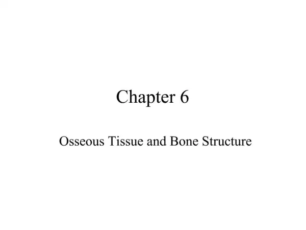
Osseous Tissue
Osseous Tissue. &. Skeletal Structures. Primary Functions of Skeletal System 1. support 2. storage of minerals & lipids -calcium salts provide vital minerals -lipids are in stored yellow marrow 3. blood cell production -RBC’s, WBC’s, and other constituents produced 4. protection

Osseous Tissue
E N D
Presentation Transcript
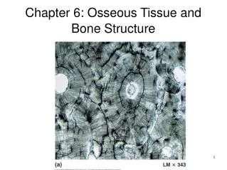
Osseous Tissue & Skeletal Structures
Primary Functions of Skeletal System 1. support 2. storage of minerals & lipids -calcium salts provide vital minerals -lipids are in stored yellow marrow 3. blood cell production -RBC’s, WBC’s, and other constituents produced 4. protection ribs: heart & lungs skull: brain vertebrae: spinal chord etc. 5.leverage: w/o bone contracting muscles just get short & fat
Classification of Bones • Every adult skeleton contains 206 bones which can be arranged into six broad categories according to shape • Long bones (relatively long and slender) • Found in arm, forearm, leg, palm, soles, fingers,toes • The femur is the largest and heaviest bone in • the body • 2. Short Bones (box-like) • Carpal bones in wrist, tarsal bones in ankle
3. Flat bones (thin roughly parallel surfaces) • Roof of skull, ribs, the sternum, scapula • Provide great protection • Provide lots of surface area for muscle attachment • 4. Irregular bones (complex shapes with short, • flat, notched or ridged surfaces) • Spinal vertebrae and several skull bones
5. Sesamoid bones (generally small, flat, and shaped • like sesame seed) • Develop inside tendons, commonly near joints • i.e.- the patellae • 6. Sutural bones (wormian bones) • Small, flat, irregularly shaped bones, form between flat bones of skull
Each bone contains two types of Osseous (bone) tissue • 1. Compact (dense) tissue: • Tightly packed • Occurs on outside surface of bone for strength & protection • 2. Spongy tissue: • Open network of struts and plates • Found in interior of the bone
Long bones • Spaces in the joints are filled with synovial fluid • The epiphysis in the joint is covered by a layer of hyaline cartilage called articular cartilage. • The medullary cavity and the open spaces of the epiphysis are filled with marrow: • a. yellow marrow: dominated by fat cells • b. red marrow: immature red, white, and blood stem cells
Flat bones • diploë contains marrow, but there is no medullary cavity • diploë is sandwiched between the internal and external table
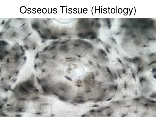
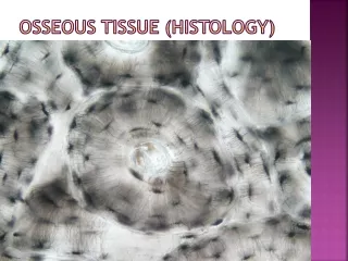
Bone Histology • Basic features of Bone organization: • matrix: very dense contains deposits of calcium • salts • 2. lacunae: pockets in the matrix which contain • osteocytes (bone cells) • 3. canaliculi: narrow passageways between lacunae • 4. periosteum: covering of bone, fiberous outer • layer, cellular inner layer
Matrix of bone • ≈2/3 of bone Ca3(PO4)2 • ≈ 1/3 of bone is collagen fibers • The rest: other Ca salts (like hydroxyapatite), ions, osteocytes and other cell types • Ca3(PO4)2 crystals are hard but brittle • They can withstand compression but will shatter when bent or twisted • Collagen fibers are very strong and flexible • Resist tension, bending, or twisting but are no good for compression
So…… hydroxyapatite crystals are tightly bound to collagen fibers giving the bone a mineral-protein composite with properties intermediate to both • Bones end up being very tough, shatter resistant, and strong. They seem to have the strength characteristics of steel reinforced concrete.
Cells in bone • There are 4 different bone cell types: • osteoprogenitor cells: stem cells which divide to • produce daughter cells which will become • osteoblasts. These are important in repairing new • bone • 2. osteoblasts: responsible for formation of new bone matrix (osteogenesis)
3. osteocytes: mature bone cells, account for most • of the bone cell population • two main functions: • a. recycle calcium salts in the matrix around them • b. participate in the repair in the of damaged • bone around them • 4. osteoclasts: giant cells, 50 or more nuclei. These • dissolve bone matrix in a process called • osteolysis releasing minerals
Compact Bone • Basic functional unit is the osteon (Haversion system) • Osteocytes are arranged around a central canal (generally run parallel to surface) • Normally this cavity contains a capillary and a venule • Other small perforating canals (Volkmann’s canals) run perpendicular to the surface • Built to withstand stress and sheer force
Spongy bone (cancellous bone) • No osteon or blood vessels • The struts and plates of the cancellous part are calledtrabeculae • Spongy bone is located at areas of low stress, or • where stress arrives from many directions • Much lighter that compact bone • The trabeculae protect the precious red marrow
Periosteum / Endosteum • All bones (except joints) are covered by a membrane called the periosteum • {fiberous outer layer, cellular inner layer} • The periosteum has three main functions: • 1. isolate bone from surrounding tissue • 2. provides route for circulatory and nervous • supply • 3. actively participate in bone growth & repair
At joints the periosteum becomes continuous with the articular cartilage, and tendons/ligaments at sites of muscle attachment • This provides for a VERY STRONG joint. • A good pull on a joint will usually break a bone before ripping these collagen fibers
A cellular layer called the endosteum covers or lines the following surfaces: • Medullary cavity • Central canals • Trabeculae • *the endosteum is active in bone growth, repair • and remodeling
Bone Growth & Development • Growth of the skeleton determines the size and proportions of our bodies • The skeleton begins to form ≈ 6 weeks after fertilization • (embryo is only 12 mm long) • Bone growth continues through adolescence with some bone continuing to grow through age 25 • Fetal skeletons are cartilaginous. They eventually turn to bone in a process called ossification
There are two types of ossification • 1. intramembranous ossification • bone develops from mesenchyme or fibrous connective tissue. • Step 1: mesenchymal cells at the ossification center secrete matrix materials (which cystalizes) and differentiate into osteoblasts • Step 2: developing bone grows outward from the • ossification center • Step 3: remodeling produces spongy & compact • bone
2. endochondrial ossification *most bone is made this way *bone replaces existing cartilage Step 1: cartilage enlarges, calcifies and dies Step 2: blood vessels grow into perichondrium Step 3: calcified cartilaginousmatrix breaks down and the fibroblasts present differentiate into osteoblasts at the primary center of ossification
Step 4: bone enlarges, medullary cavity is formed, bone increases in length and diameter Step 5: center of epiphysis begin to calcify, secondary ossification centers arise at the ends of major long bones Step 6: epiphyses filled with spongy bone and covered with hyaline cartilage
The Blood and Nerve Supply • Bones of the skeleton typically have an extensive blood supply • 3 major sets of blood vessels develop • The nutrient artery and vein: • Supply the diaphysis / enter through one or more foramina • 2. Metaphyseal vessels: • Supply blood to the epiphyseal plate where bone growth is most rapid
3. Periosteal vessels: • Supply surface cells of bone, develop in from periosteum • *Eventually, as the bone hardens all three of these supplies interconnect • Sensory nerves enter with the nutrient artery and form a large nerve network. Injuries to bone tend to be very painful
Bone is a very Dynamic Tissue • As part of a bone maintenance program, your bones are being broken and “reproduced” throughout life • Involves a series of metabolic activities between osteoblasts, osteoclasts, and osteocytes • Generally the activity between these cell types is balanced. As one osteon is produced, one is destroyed, etc. • Turnover rate is high. In young adults 1/5 of skeleton is replaced each year
There are regional differences • Spongy bone in head of femur is replaced 2-3 times/year • The compact bone in the dyaphysis remains largely unchanged • Effects of exercise on bones • Bones adapt to new stresses
Theory for mechanism • Stressing bone generates minute electric fields around mineral crystals • Osteoblasts are apparently attracted to the electric field • Once in the area they begin to produce bone • Electrical fields are also used to stimulate the repair of bones in severe fractures
Because bones are adaptable they reflect the forces and stresses applied to them • Heavily stressed bones become thicker and stronger • Bones subject to ordinary stress become thin and brittle
Normal bone growth depends on nutritional and hormonal factors 1. The body must have a constant supply of dietary calcium and potassium salts. Also required in trace amounts are: Mg, Fe, Fluoride, and Mn 2. Calcitrol is a hormone produced in the kidneys and is essential for normal Ca+2 & K+ ion absorption in the digestive tract. 3. Vitamin C must be available in the diet 4. Vitamin A, K, & B12 also have a significant effect on bone development
5. Growth Hormone from the pituitary gland & Thyroxin from the thyroid gland stimulate bone growth 6. Sex hormones estrogen and androgen stimulate bone growth 7. parathyroid hormone: ↑ calcium ion conc. In body fluids 8. calcitonin: ↓ calcium ion conc. In body fluids
Fracture Repair • Most bone fracture or breaks will repair (even after severe damage) if the blood supply is not interrupted