Download
1 / 59
590 likes | 699 Vues
Explore the differences between asexual and sexual reproduction, including mechanisms, patterns, and advantages/disadvantages. Learn about unique processes like parthenogenesis and hermaphroditism, and understand the male reproductive system and spermatogenesis in detail. Discover how hormones impact spermatogenesis and male secondary sex characteristics.

E N D
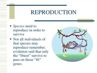
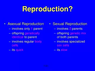
Asexual versus Sexual Reproduction Asexual reproduction involves only one parent and results in the production of clones. Different mechanisms include: fragmentation, budding, gemmule formation, regeneration, and parthenogenesis. Sexual reproduction involves two parents (each producing, in general, a haploid gamete) and the union of these gametes to form, in general, a diploid offspring. Some animals alternate between the two types. Each mechanism has its pros and its cons.
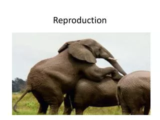
Various patterns of Sexual Reproduction Hermaphroditism Parthenogenesis Sonoran Spotted Whiptails are all female and reproduce via parthenogenesis (virgin birth). A Sonoran Spotted Whiptail's eggs contain complete copies of her genome and will develop into embryos despite not being fertilized by a male. The resulting female offspring are all clones of their mother. While their reproduction is asexual, these parthenogenic lizards do exhibit sexual behavior and will sexually interact with each other depending on their fertility cycle
Sequential Hermaphrodite - an organism with both male and female sex organs that mature at different times Protandry - where an organism is born male and changes into a female. As in protogyny, if the female is removed from the group, the dominant male begins the transition from male to female and the next-dominant male becomes the breeding male in the “harem”. Protogyny - where an organism is born female and changes into a male. If the dominant male is removed – either physically removed or if it dies for some reason – the largest, most dominant female in the group is triggered to become male and take over control of the harem
External fertilization Involves the shedding of gametes outside of the body. Involves the absence of copulatory structures or other specialized structures for internal fertilization Fertilization occurs outside of the body. External fertilization occurs in many aquatic animals (many fish, most amphibians, sea urchins etc.)
Internal fertilization Involves the deposition of sperm by the male (actively or passively) into the female. Involves copulatory structures (e.g., claspers, penis) or specialized structures (e.g., spermatophores) or positioning of cloacas next to each other (e.g., birds). Occurs in many terrestrial organisms and some aquatic organisms ranging from invertebrates to mammals.
Testes (within scrotum) The scrotum is a sac that contains the testes (sperm production requires temperatures around 34 C). The testes (or singular testis) or testicles are the paired gonads
The structure of the testes The coiled seminiferous tubules are the site of spermatogenesis (see below) Within the seminiferous tubules are the spermatogenic cells, spermatogonia, and primary and secondary spermatocytes, spermatids, and sperm Also within the seminiferous tubules are the sustentacular cells (AKA Sertoli cells) that extend from the basement membrane to the lumen of the tubule. In the spaces between adjacent seminiferous tubules are clusters of interstitial endocrinocytes of Leydig cells that secrete testosterone.
Spermatogenesis takes about 74 days It begins in the spermatogonia which are stem cells undergo mitosis Some of the daughter cells of the spermatogonia differentiate into primary spermatocytes (2n) Before dividing, the primary spermatocytes enlarge. They then go through Meiosis I during which crossing-over occurs (resulting in an increase in genetic variability). The products of Meiosis I are the secondary spermatocytes (each with 23 chromosomes and thus haploid but sister chromatids still are present) Meiosis II results in spermatids (each with 23 chromosomes and only one chromatid) The final stage is called spermiogenesis and involves the maturation of spermatids into sperm that mature at about a rate of 300 million/day. Each sperm is composed of a head (containing the DNA and a lysosome-like structure the acrosome which contains enzymes that penetrate the oocyte), a midpiece which carries on metabolic activities, and a tail which is a flagellum for propelling the sperm along.
Hormones involved in spermatogenesis and male 2o sex characteristics Follicle Stimulating Hormone (FSH) and Leutinizing Hormone (LH) from the Anterior Pituitary (both stimulated by GnRH) play roles in spermatogenesis. LH stimulates the Leydig cells to produce testosterone. FSH acts indirectly in that FSH and testosterone act synergistically on the sustentacular cells to stimulate the secretion of androgen-binding protein (ABP) which binds testosterone and thus keeps the levels of testosterone high around the seminiferous tubules. Testosterone then stimulates the final steps of spermatogenesis. Androgens (including testosterone) are also involved in the development of the male reproductive system, sexual characteristics, libido, and metabolism.
Ducts of the male reproductive system Part I The various ducts play roles such as sperm storage, propulsion, and maturation (epididymis), sperm transport (straight tubules, vas deferens, ejaculatory ducts, and urethra. The ducts of the testes lead to the epididymis. The epididymis is a comma-shaped organ that lies along the posterior border of each testis
Ducts of the male reproductive system Part II The vas deferens ascends along the posterior border of the epididymis and penetrate the inguinal canal and then the pelvic cavity. The ejaculatory ducts lie posterior to the urinary bladder. The urethra is the shared passageway of the reproductive and urinary systems.
Glands of the male reproductive system Glands function in the production of semen that is a mixture of sperm and seminal fluid. The normal volume is 2.5-5 ml. with a sperm count of 50-150 million sperm/ml.
The seminal vesicles secrete an alkaline (for the neutralization of acidic fluids in the female reproductive tract), viscous fluid that contains fructose (ATP production), prostaglandins (sperm motility and viability), and clotting proteins which make up about 60% of the volume of the semen.
The prostate gland secretes a milky, slightly acidic fluid that contains citrate (for ATP production), acid phosphatase (unknown), and proteolytic enzymes. These secretions make up about 25% of the volume of the semen.
The bulbourethral glands secrete an alkaline fluid and mucus that neutralize acids in the urethra and decrease damage to sperm, respectively.
Oogenesis I The female gonads are the paired ovaries (sites of oogenesis) Ovarian follicles consist of oocytes in various stages of development. The most immature of the cells are surrounded by a single layer of follicular cells, but as they mature they are surrounded by several layers which are referred to as granulosa cells (these cells nourish the developing oocyte as begin to secrete estrogen as the follicle grows). A mature or Graafian follicle is a large fluid-filled follicle that ruptures and expels a secondary oocyte during ovulation.
Oogenesis II A corpus luteum is the remnant of an ovulated mature follicle. It produces progesterone, estrogens, relaxin, and inhibin until it degenerates into a corpus albicans (white body). Oogenesis like spermatogenesis involves two reductional divisions (Meiosis I and II). During early fetal development the primitive germ cells differentiate into oogonia which are diploid. These cells divide mitotically producing millions of germ cells. However, many of these cells will degenerate (atresia). A few will develop into primary oocytes that enter but do not complete Meiosis I until after puberty. At birth there will be 200,000-2,000,000 oogonia and primary oocytes in each ovary. However, only about 400 of these will mature and ovulate, the others undergo atresia. As a follicle grows additional layers of cells surround it. A clear glycoprotein layer, the zonapellucida will form between the oocyte and the granulosa cells. The innermost layer of granulosa cells is called the corona radiata. The cavity is the antrum. A follicle containing these multiple layers and an antrum is called a secondary follicle.
Oogenesis III After puberty, LH and FSH stimulate one secondary follicle per month to resume meiosis. The oocyte then completes meiosis I producing one secondary oocyte and one polar body of unequal sizes. The secondary oocyte then enters Meiosis II but is arrested in metaphase. This is the mature or Graafian follicle. At ovulation, the secondary oocyte is expelled into the pelvic cavity and usually swept into the Fallopian tube. If fertilization does not occur, it degenerates. But if fertilization does occur meiosis II resumes. Again there are two cells of unequal size produced. One is the ovum (the larger cell) and the other is the second polar body. When the nuclei of the sperm and ovum unite a zygote is formed
The uterine or fallopian tubes are paired structures that extend laterally from the uterus.. Fertilization occurs in the Fallopian tubes which serve as a passageway for the secondary oocyte or zygote.
The uterus (AKA womb) is a pear shaped organ. The layers of the uterus include: an outer perimetrium or serosa, a middle muscular myometrium consisting of three layers of smooth muscle fibers, and an inner endometrium which is highly vascularized and divided into two layers: the stratum functionalis which is shed during menstruation and the deeper stratum basalis which is permanent. The uterus serves as a pathway for sperm, the site of menstruation, the site of implantation, and the site of development of the fetus.
The vagina is a tubular, fibromuscular organ lined with mucous membrane that is continuous with the uterine mucosa. The muscularis is composed of smooth muscle. The vagina serves a passageway for menstrual flow, it receives semen, and it is a passageway for childbirth.
The mammary glands are modified sudoriferous (sweat) glands that produce milk.
Osmoregulation and Excretion
Water Balance and Excretion. Water balance is necessary for the maintenance of a steady state. Mechanisms differ but the excretory system is usually involved.
Basic problems I getting water or losing water and the maintenance of solutes that function in water balance 1. In mammals for example, water is gained from absorption from solid food and from liquids, as well as from metabolic processes. 2. In mammals, for example, water is lost by: urinary excretion, evaporation from the lungs and skin, sweating, and elimination in the feces.
Basic Problems II 3. Solutes are gained by: absorption from food and from liquids, secretion, cellular respiration, and metabolism. In aquatic animals it occurs by osmosis from the environment. 4. Solutes are lost by: urinary excretion, respiration, defecation, and sweating. In aquatic animals it also occurs by osmosis.
Osmoconformers versus osmoregulators Note that stenohaline refers to animals that cannot tolerate substantial changes in external osmolarity, whereas euryhaline animals can survive radical fluctuations.
Osmoconformers are organisms that do not actively adjust their internal osmolarity relative to their changing environment. Many marine invertebrates are osmoconformers. Sharks are also, but they are not the best osmoconformers and they must use energy to maintain osmotic gradients and maintain higher levels of urea. Sharks possess a salt-secreting organ, the rectal gland. Hagfishes (see pic) are osmoconformers
Osmoregulators are organisms that maintain an internal solute concentration that does not vary regardless of the external environment.
marine organisms who live in an environment where their surroundings contain more solutes than the bodies of the organisms. Thus, they have a problem with water loss. They drink lots of water and pump salt out.
freshwater organisms live in an environment where their surroundings contain fewer solutes. Thus, water moves into the animals, and in response many of them produce copious amounts of urine and absorb salts in food and actively uptake it from their surroundings.
Animals that live in ephemeral bodies of water must adapt to periodic dry conditions
terrestrial organisms have a problem with desiccation, thus they possess cuticles or barriers to water loss. Some animals can be dormant for long periods of time (anhydrobiosis). They also replenish lost water or conserve water (e.g., behaviorally).
Protonephridia are the simplest type (the flame-cell system of flatworms but also found in other organisms). Interstitial fluid is filtered across the membranes of these cells.
Metanephridia consist of tubules that drain to the outside through a nephridiopore after filtration and occur in most annelids.
Malpighian tubules remove nitrogenous wastes in insects and other terrestrial vertebrates.
Vertebrate (e.g., human) excretory system Structures and functions
Kidney Anatomy the deep vertical fissure near the center of the concave border is the renal hilus through which the ureter, blood vessels, lymphatic vessels, and nerves leave the kidneys the renal cortex if the superficial layer of the kidneys the medulla is the inner region within the medullar are 8-18 cone-shaped renal (medullary) pyramids the apex of each pyramid is the renal papilla the portions of the cortex that extend between the pyramids are the renal columns the cortex and pyramids make up the parenchyma which houses the nephrons (the functional units of the kidney) the ureter expands within the kidney forming the renal pelvis which is subdivided into major and minor calyces