Highly Conserved Structures in PTP1B Domain
10 likes | 116 Vues
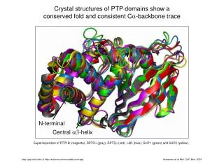
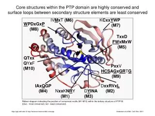
Explore core structures within the PTP domain, with conserved motifs highlighted on a ribbon diagram of PTP1B's tertiary structure. Learn more about this conservation and surface loops. Published in Mol. Cell. Biol. 2001.

Highly Conserved Structures in PTP1B Domain
E N D
Presentation Transcript
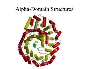
IVMxT (M6) KCxxYWP (M7) WPDxGxP (M8) TxxD FWxMxW (M5) QTxx QYxF (M10) PxxV HCSAGxGRTG (M9) IAxQGP (M4) DxxRVxL (M2) NxxKNRY (M1) DYINA (M3) Core structures within the PTP domain are highly conserved and surface loops between secondary structure elements are least conserved Ribbon diagram indicating the position of conserved motifs (M1-M10) within the tertiary structure of PTP1B (blue - most conserved; red - least conserved). http://ptp.cshl.edu & http://science.novonordisk.com/ptp Andersen et al Mol. Cell. Biol. 2001