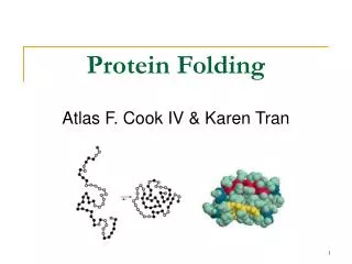
Protein Structure and Folding Simulation
This review explores the hierarchical organization of protein structures starting from amino acids. It explains how the primary structure consists of peptide bonds formed through dehydration synthesis, leading to various configurations. The secondary structures, such as alpha helices and beta-pleated sheets, are quantified by hydrogen bonds, while tertiary structures arise from interactions of R groups, exemplified by sulfur-sulfur bonds. Finally, the formation of quaternary structure from multiple polypeptide chains is discussed, emphasizing the importance of structural integrity and genetic conditions affecting protein functionality.

Protein Structure and Folding Simulation
E N D
Presentation Transcript
Amino Acid Review • All amino acids have a common structure, but differ in their R groups. • The asymmetric carbon must have the four attachments in a particular order or isomers will be formed
Primary Structure • By holding hands you simulate a “dehydration synthesis reaction” by linking the amine group of one amino acid to the carboxyl of the second amino acid. The holding of hands represents the “peptide bond”. Points to understand: • The amino acids (students) can be arranged in any order. Each different arrangement produces a new protein. • The protein has a free amine end and a free carboxyl end (students at each end with a free hand). • The structure is flexible and can bend and fold. • A typical polypeptide or protein often has hundreds of amino acids.
Secondary Structure • The line of students is bent so that it forms a “U” shape with one end of the line across from the other end. The students formed a “beta pleated sheet” structure from this configuration. By alternating with their arms “up” or “down” to create the pleated sheet. One side of the pleat (arms up or arms down) can stretch across the gap between the two lines so that the lines come together at these points. Hydrogen bonds between the two lines can be formed by extending out the thumbs (or other fingers) and “hooking” with the counterpart on the other line.
Secondary Beta Pleated Sheet So arms up on one side were the double bonded C from the carboxyl end and the arms down were the hydrogen from the other sides amine group (the up and down are not relevant – just for the simulation).
Secondary Structure Points to understand: • The Hydrogen bonds form at regular intervals, between the backbone components. • The Hydrogen bonds are “weak” compared to the peptide bonds. *The number of the H bonds in the entire structure does give strength to the structure (so alone H are weak, a lot of them are stronger) • The Hydrogen bonds and the pleats create a regular structure from the amino acid chain. • Note–can’t construct alpha helixes using students, but can you visualize this structure?
Secondary Structure Alpha Helix Alpha helix Note this is not showing R groups
Tertiary Structure • The line of students (primary structure) is bent at some point so that two amino acids (students) are physically near each other. Asulfur-sulfur bond between these two students was created using a bungee cord. The “S” hooks on the ends of the bungee cord hooked through belt loops to join these two amino acids together. Points to understand: • The “S” shaped hooks on the bungee cord represent sulfur atoms. The tertiary structure is formed when two sulfur atoms are joined bonded together by the strong non-polar covalent bond. • S-S bonds form from the interactions between the side chains – “R” groups. • The S-S bond is more rigid than Hydrogen bonds. This gives the protein structure more rigidity and stability. • The size of the loops created by the S-S bonds depends on the location of the sulfur containing amino acids (primary structure).
Quaternary Structure • Form two or more small polypeptide chains. The chains can be positioned side by side or in other configurations. A quaternary structure is formed when two or more polypeptides come together to make the functional protein. Points to understand: • Two or more separate polypeptides are needed for this level of protein structure. • Each polypeptide has its own primary, secondary and tertiary structure. Often these levels of structures provide the fit between the different polypeptides in forming the functional protein. • Genetic conditions such as Thalasemia occur when the polypeptide units are not produced in equal amounts. The result is that one polypeptide chain lacks a sufficient number of partners to form the final functional protein unit.