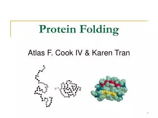
Protein FOLDING
Protein FOLDING. Proteins are composed of amino acids linked with peptide bond The way or sequence b y different amino acids are linked together makes the primary structure of a protein. . proteins. Peptide bond formation. This polymerization of amino acids is what creates proteins.


Protein FOLDING
E N D
Presentation Transcript
Proteins are composed of amino acids linked with peptide bond The way or sequence b y different amino acids are linked together makes the primary structure of a protein. proteins
Peptide bond formation This polymerization of amino acids is what creates proteins. This condensation reaction yields the newly formed peptide bond and a molecule of water.
Properties of Peptide bond Joins amino acids • 40% double bond character • Caused by resonance • Results in shorter bond length • Double bond disallows rotation
Biochemist’s peptide unit – from N to C – all main chain atoms within the unit lie in the same residue Structural biologist’s peptide unit – from Cα to next Cα - all main chain atoms within the unit lie in a plane
Torsion Angles • A torsion angles is defined by 4 atoms; • the carboxylic carbon, C1 • the alpha carbon, C2 or C-alpha; and the amide group nitrogen, N. • THREE repeating torsion angles along the peptide backbone chain are; • phi (φ) N-CaSingle bond • psi (ψ) Ca-COSingle bond • omega (ω) C-NPartial double bond Dihedral angle— A measure of the rotation about a bond, usually taken to lie between -180° and +180°. Dihedral angles are sometimes called torsion angles
Planar Peptide bond has resonance structures • Peptide bond has resonance structures --> partial double bond character • Peptide bond is a partial double bond, so backbone can only rotate around Psi (ψ) and phi (φ) bonds.
The OMEGA angle tends to be planar (0 or 180o) due to delocalization of the carbonyl pi electrons & the nitrogen lone pair. Due to the partial double bond character of the peptide bond; six atoms (Cα, C, O, N, H, and Cα) lie in a plane in a pair of linked amino acids - there is no rotation about the peptide bond (peptide). Peptide Bonds Are Planar
Trans is generally favored over cis: Generally, the two Cα groups in a trans configuration experience minimum steric interaction (cis/trans) than when in cis. Because of the partly double nature of the peptide bond, ω is always close to 180o for trans- peptides or 0o for cis- peptides (±30o in exterme cases) Cis- peptides are energetically extremely unfavourable (~1000 fold) because of steric clashes between the neighbouring Cα atoms The trans configuration is adopted for almost all peptide bonds
(phi) is limited Proline’s Cyclic Structure is Particularly Limiting some specific bonds are often cis • The only exception is peptide bond before proline, where cis- peptide is just 4 times less favourable than trans- peptide, because there are some steric clashes in both • cis- and trans- forms • Tyr-Pro (25%), Ser-Pro (11%), X-Pro (6.5%)
Proline adopts cis and trans forms • Proline cis-trans isomerization is an important factor in protein folding – prolylpeptydyl isomerases catalyzeisomerization step • According to statistics, 0.03% of non-proline peptides and 5.2% of X-Pro peptides are in cis- conformation, resulting in a total of 0.3 % cis-peptides. It’s in cis configuration (6% of the time), frequently in b-turns
The peptide backbone conformations can be described by Phi (φ) and Psi (ψ), pure single bonds. The two adjacent rigid peptide units may rotate about these bonds, taking on various orientations. This freedom of rotation about two bonds of each amino acid allows proteins to fold in many different ways. The key to protein folding lies in the torsion angles of the backbone
The peptide bond constrains the polypeptide The length and angles are fairly invariant in the known protein structures The planarity of the peptide bond restricts omega to 180 degrees in very nearly all of the main chain peptide bonds. In rare cases omega = 0 degrees for a cis peptide bond which, usually involves proline. • φ and ψ are flexible, therefore rotation occurs here • However, φ and ψ of a given amino acid residue are limited due to steric hindrance like clashes of backbone atoms and Cb atom • Only 10% of the {φ, ψ} combinations are generally observed for proteins • First noticed by G.N. Ramachandran
Main Chain Conformations Factors that influence the conformational equilibria of peptide chains are: • Conformations are defined by dihedral angles Φ & Ψ. • Hydrogen bonding of amide carbonyl groups to N-H donors. • Steric crowding of neighboring groups. • Repulsion and attraction of charged groups. • The hydrophilic and hydrophobic character of substituent groups. Interference to rotation caused by spatial arrangement of atoms within molecule • Atoms cannot overlap • Atom size defined by van der Waals radii • Electron clouds repel each other
Steric Collisions in addition to peptide bond φ = 0 ψ = 0 combination is forbidden Collision comes from peptide backbone and from R group.
Alpha-helix Secondary Structure Elements
Secondary structure elements in proteins A secondary structure element is a contiguous region of a protein sequence characterized by a repeating pattern of main-chain hydrogen bonds and backbone phi/psi angles alpha-helix (local interactions) beta-strand(non-local interactions)
Elements of protein structure 2˚ 1˚ Primary structure - the covalent Structure of the protein: sequence, Plus disulfides and modifications. Secondary structure - Local conformation of some part of a polypeptide. Primary structure determines secondary structure -
Secondary Structure • Can a polypeptide chain fold into a regularly repeating structure? • In 1951, Linus Pauling and Robert Corey proposed two periodic structures called the α helix (alpha helix) and the β pleated sheet (beta pleated sheet). • Subsequently, other structures such as the β turn and omega (Ω) loop were identified. • Protein 2ndary structure takes one of three forms: • α helix • β sheet • Turn, coilor loop • 2ndary structure are tightly packed in the protein core in a hydrophobic environment • Although not periodic, these common turn or loop structures are well defined and contribute with α helices and β sheets to form the final protein structure.
The reasons of secondary structure formation • Since sidechains of hydrophobic residues are located in the hydrophobic core, the mainchain atoms of the same residues in most cases are also within the hydrophobic core. • Since the presence of polar groups in hydrophobic environment is very unfavourable, the main chain N- and O- atoms have to be neutralised by formation of hydrogen bonds. • The two most efficient ways of hydrogen bond formation is to build an alpha helix or a beta sheet Reflects the tendency of backbone to hydrogen bond with itself in a semi-ordered fashion when compacted
the main-chain can hydrogen bond to itself there are also side-chain acceptors and donors the carbonyl oxygen: main-chain hydrogen bond acceptor the amide nitrogen: main-chain hydrogen bond donor
Main-chain H-bonds make proteins special “It would seem extraordinary that no other polymer structures exist in which internal hydrogen bonding can give rise to periodically ordered conformations, but no others have been found thus far. We are therefore forced to recognize the uniqueness of this capacity in polypeptide chains, one which enables them to meet the exacting and sophisticated demands of structure and function” --Doty P, Gratzer WB in Polyamino acids, polypeptides and proteins, pp. 111-118, 1962, University of Wisconsin Press • 2° structure full of H-bonds • Why so much 2° structure? • Saturate with H-bonds • neutralize dipoles in buried regions. • Provide rigidity, stability: • let mutations change chemical properties only • not conformation.
The Alpha Helix • Figure 3.29. Structure of the α Helix. (A) A ribbon depiction with the α-carbon atoms and side chains (green) shown. (B) A side view of a ball-and-stick version depicts the hydrogen bonds (dashed lines) between NH and CO groups. (C) An end view shows the coiled backbone as the inside of the helix and the side chains (green) projecting outward. (D) A space-filling view of part C shows the tightly packed interior core of the helix.
The Alpha Helix • Most common (about 25% of the amino acids in proteins are in this structure) • Repetitive secondary structure • 3.6 residues per turn; pitch (rise per turn) = 5.4 Å • C′=O of i forms H bonds with NH of residue i+4 • Intra-strand H bonding • C′=O groups are parallel to the axis; side chains point away from the axis • All NH and C′O are H-bonded, except first NH and last C′O • H-bonds are in-line, optimum distance • Hence, polar ends; present at surfaces • Right –handed favored due to steric constrains • Stability is greatly enhanced by internal van der Waals contacts • R-groups project outward, and provide the main constraints on helical structure • Interactions do not involve side chains • Interaction with other helices, charged chains, ions and molecules
The Alpha Helix Is a Coiled Structure Stabilized by Intra-chain Hydrogen Bonds • The α helix is stabilized by hydrogen bonds between the NH and CO groups of the main chain…. situated four residues ahead in the sequence. • Dipoles of H-bonding atoms NH and CO are well aligned along the axis of helix. • Except for amino acids near the ends of an α helix, all the main-chain CO and NH groups are hydrogen bonded. • Each residue rises 1.5 Å along the helix axis…a rotation of 100 degrees…3.6 amino acid residues per turn of helix. pitch of 5.4 Å. • Amino acids spaced three and four apart in the sequence are spatially quite close to one another in an α helix. • Radius allows favorable van der Waals contacts across helical axis. • Side chains are well staggered minimizing steric interference. • The internal cavity is dominated by H-bonds and (all atoms at allowed intra-atomic contacts).
α-Helix has: • 3.6 residues per turn • Rise/residue = 1.5 Å • Rise/turn = 5.4Å • Hydrogenbonds are made between residues n and n+4
Hydrogen-Bonding Scheme For an α helix In the α helix, the CO group of residue n forms a hydrogen bond with the NH group of residue n+ 4.
Dipole moment • A helix has an overall macro dipole moment , -ive at carboxy end and +ive at amino end. • Helix dipole results from orientation of CO - NH hydrogen bonds of the peptide bond pointing along the helix axis. • The dipole is neutralized by H-bonds between NH and CO groups in the center of helix but ends. • The overall dipole can lead to destabilization of the helix through entropic effects. • As a result, α helices are often capped at the N-terminal end by a negatively charged amino acid, such as glutamic acid, in order to neutralize this helix dipole. • Less common (and less effective) is C-terminal capping with a positively charged amino acid, such as lysine.
The dipole moment of a helix The N-terminal positive charge is commonly used to bind negatively charged ligands such as phosphate groups, because the backbone amides can serve as hydrogen bond donors.
3.10 helix pi-helix Other Types of Helical Structures
Secondary Structure – Triple Helix • Three polypeptide chains woven together • Glycine, proline, hydroxy proline and hydroxylysine • H bonding between –OH groups gives a strong structure • Typical of collagen, connective tissue, skin, tendons, and cartilage
R-groups extend radially from the α-helix core, shown in helical wheel diagram. α -helices can be: Polar Amphipathic Hydrophobic
Amino-acid propensities Different amino-acid sequences have different propensities for forming α-helical structure. • Methionine, alanine, leucine, glutamate, and lysine ("MALEK“ ) all have especially high helix-forming propensities. • Proline, glycine, tyrosine and serine have poor helix-forming propensities. • Proline tends to break or kink helices because it cannot donate an amide hydrogen bond (having no amide hydrogen), and because its side-chain interferes sterically. • Proline’s ring structure also restricts its backbone φ dihedral angle to the vicinity of -70°, which is less common in α-helices. • However, proline is often seen as the first residue of a helix, presumably due to its structural rigidity. • At the other extreme, glycine also tends to disrupt helices .. its high conformational flexibility makes it entropically expensive to adopt the relatively constrained α-helical structure.
Five different kinds of constraints affect thestability of an α-helix: • Electrostatic repulsion (or attraction) between successive amino acid residues with charged R groups (2) Bulkiness of adjacent R groups (3) Interactions between R groups spaced three (or four) residues apart (4) Occurrence of Pro and Gly residues (5) Interaction between amino acid residues at the ends of the helical segment and the electric dipole inherent to the α- helix. The tendency of a given segment of a polypeptide chain to fold up as an α-helix therefore depends on the identity and sequence of amino acid residues within that segment.
Hydrophilic Hydrophobic Globular protein. Where are -helices? • Hydrophobic helices cross lipid membranes. • Amphiphilic are common: • Surfaces of globular proteins. • Membrane surface proteins, e.g. mellittin
Beta Pleated Sheet Secondary Structure
The β sheet (also β-pleated sheet) is the second form of regular secondary structure in proteins. A single stretch of β-structure is termed a β-strand. The sheets are made of individual beta strands - The first β sheet structure was proposed by William Astbury in the 1930s. - A refined version was proposed by Linus Pauling and Robert Corey in 1951. β sheet
The β-strand • A beta strand (also β-strand) is a stretch of amino acids; • Typically 5–10 amino acids long. • Peptide backbones are almost fully extended. • Highly extended form of polypeptide chain. • Adjacent side chains point in opposite directions. • A beta strand usually is associated with other beta strands…. sheet
Beta Sheet Conformation consists of; Several β-strands associate laterally to form β-sheets. Sheets are stabilized by backbone hydrogen bonds. Sheets can be made of nearby (in sequence) or faraway strands. Main chain H-bonding between CO and NH on adjacent strands is a major contribution to stability R groups extend above and below the sheet…. along each strand, they alternate in pointing “up” and “down” Beta-sheets are very common in globular proteins mostly less than six strands. With a width of approximately 25 Ǻ No preference for parallel or antiparallel beta-sheets • Beta strands often interact to form beta sheets: • - Parallel • - Antiparallel: optimum H-bonding • - Mixed
Unlike α-helices: • Are formed with different parts of the sequence • H-bonding is inter-strand (opposed to intra-strand) • Side chains from adjacent residues on opposite sides of the sheet and do not interact with one another. • Peptide groups of adjacent residues point in opposite directions. • The axial distance between adjacent residues is 3.5 Ǻ (0.35nm). • Two residues per repeat unit which gives the beta-strand a 7 Ǻ pitch. • In alpha-helix the axial distance between adjacent residues is only 1.5 Ǻ. • Polypeptides are far more extended than those in the alpha-helix. Sheets are common in the core of protein structures Like α-helices: • Repeating secondary structure (2 residues per turn) • Can be amphipathic β−sheets vs. α-helices β strands are arranged adjacent to other strands and form an extensive hydrogen bond network with each other. N-H groups in the backbone of one strand establish hydrogen bonds with the C=O groups in the backbone of the adjacent strands.
A mixed b sheet: strand directions • An individual strand may exhibit a mixed bonding pattern, with a parallel strand on one side and an antiparallel strand on the other. • Mixed sheets are far less common indicating that this pattern is less stable than the antiparallel arrangement.
The "pleated" appearance of β strands arises from……. tetrahedral chemical bonding at the Cα atoms of the peptide bond which alternates above and below the plane of the sheet. . Pleating causes the distance between adjacent Cα atoms 6Å ….7.6Å (2 × 3.8 Å) expected from two fully extended trans peptide virtual bonds. “Sideways" distance between adjacent Cα atoms in H-bonded β strands is roughly 5 Å. β -Sheets are said to be pleated
0.7 nm Pleated antiparallel sheet Antiparallel • Note: • H-bonds • R-groups orientation, distance. • Pleated character • Ave. strand length is about 6 aa’s
0.65 nm Pleated parallel sheet Parallel • Note: • H-bonds not at optimal angle. • R-group orientations, distances • Pleated character • Avg. Strand length ~ 6 aa.