Download
1 / 83
830 likes | 979 Vues
Digestive Tract: Let’s Get to the Bottom of it. By: Diana Blum RN MSN Metropolitan Community College. Primary Role. Extract molecules essential for cellular function from fluids and food. Ingestion, Digestion, Absorption, Elimination.

E N D
Digestive Tract: Let’s Get to the Bottom of it By: Diana Blum RN MSN Metropolitan Community College
Primary Role • Extract molecules essential for cellular function from fluids and food.
Ingestion, Digestion, Absorption, Elimination • Digestion: breakdown of food into simple nutrient molecules that can be used by cells • Process requires: • 1. • 2. • 3. http://health.discovery.com/centers/digestive/machine.html
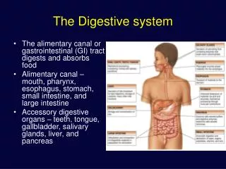
Digestive tract • Also called ___________tract • muscular tube about 30 ft long • Main parts • Mouth • Pharynx • Esophagus • Stomach • Small intestine • Large intestine • Anus
Acessory Organs • Salivary glands • Liver • Gallbladder • Pancreas • Each of the above accessory organs secrete fluid that contain special enzymes that enable breakdown (metabolism) of food • Peritoneum lines the abdominal cavity and covers surface of organs • Enables organs to moves without friction during breathing and digestion
mouth • Teeth cut and grind food • Salivary glands secrete saliva • Saliva: • Amylase: • Tongue mixes saliva with food and when small enough- forces the food into the pharynx
Pharynx • Shared by digestive and respiratory tracts • Joins mouth and nasal passages • Contains the epiglottis • Covers the airway (like a trap door) to prevent food from entering respiratory tract
esophagus • Long muscular tube that passes through the diaphragm into the stomach • Gravity helps move the food but it is not essential • Circular, wave like contractions of the muscles propel food down the tract (peristalsis)
Stomach • Widest section of the GI tract • Separated from esophagus by the cardiac sphincter • Has 3 sections • Unique muscle layers churn food by mixing it with gastric secretions • Rennin-starts breakdown of milk proteins • Pepsin-partially digests protein • HCL acid-partially digests protein • Lipase-breaks down fat • Chyme: • Pyloric sphincter- keeps food in stomach until it is mixed properly
Small Intestine • Chyme leaves stomach and enters here • Chemical digestion and absorption of nutrients take place • 20 feet long • 3 sections • Duodenum-liver and pancreatic enzymes enter here • Jejunum • Ileum
Small Intestine Continued • Bile- produced in the liver and stored in the GB break down large fat globs • Pancreatic enzymes-reduce the fat to glycerol and fatty acids to be easily absorbed • 3 layers of tissue make up the wall • Mucous membrane-secretes digestive enzymes • Sucrase, lactase, maltase, lipase, etc. (see table 36-1) • Inner layer- covered with Villi (microscopic projections). Digestive food molecules are absorbed through the villi into the bloodstream • Muscle layers continue to contract moving the chyme into the large intestine.
Large Intestine • No Villi • No digestive enzymes • Chyme enters through the ileocecal valve • Water is absorbed and remaining waste=feces • 5 sections • Cecum-1st section..appendix is here • Ascending colon-up right abdomen • Transverse colon- across abdomen just below waist • Descending colon-down the left abdomen • Sigmoid colon-the part of the descending colon between iliac crest and rectum • Rectum-the last 6-8 inches of the large intestine • Anus – where waste leaves the body
Age related changes • Teeth mechanically worn down • Illness causes increased risk for problems with digestion/elimination • Gingiva recedes • Tooth loss from caries and periodontal disease • Loss of taste buds • Xerostomia (dry mouth) is common • Walls of esophagus and stomach are thinner with lessened secretions • HCL Acid and digestive enzyme production decreases • Gastric motor activity slows • Delayed gastric emptying • Hunger contractions diminish • In the large intestine- muscle layer and mucosa atrophy • Smooth muscle tone and blood flow decreases • Connective tissue increases • Constipation is frequent • More laxative use
Nursing Assessment • Hx of illness: weight loss, indigestion, change in bowel habit • PMH: surgery, trauma, infection, burns, hepatitis, ulcers, cancer, stomas, meds, allergies • Fam Hx: diabetes, CA, ETOH, polyps, obesity, ulcers, GB Dx • System Review: flatus, dyspepsia (indigestion), skin changes, caries, diff chewing, abd distention, pain, elimination • Functional: nutrition, activity, meal times, likes/dislikes, food beliefs • Physical exam: mucous membranes, condition of mouth/teeth, abd distention, bowel tones, palpation, percussion, rectum/anus for lesions, color, hemorrhoids
diagnostics • Imaging/radiographs: NPO, allergy (iodine, dye, shellfish), consent • UGI • Barium swallow/enema • Endoscope • Upper • Lower • Hemmocult-looks for blood
NG Salem Sump
Tube feedings • Assist pt into fowlers to reduce aspiration. • Remains this way for 30 minutes after • Pt remains up at least 30 degree during continuous feeding • Check placement for tube in stomach or duodenum prior to use • Air bolus and residual • Check to make sure you have the correct formula • Stop feeding if nausea or pain • Rinse tube with 30 cc fluid after each bolus • Administration • Remove plunger • Pinch tube while inserting syringe to avoid stomach content leak • Hold barrel about 12 inches above stomach and allow gravity to infuse • Flush after bolus complete
GI decompression • Ng with suction • removes fluid and gas • To use • Attach to sxn as ordered • Generally low, intermittent is used for single tube • Low continuous for dual lumen tubes • Check patency • Irrigate routinely • Monitor output • Assess for flatus • Provide comfort measures • Once tube in place- securely tape it to nose
TPN Deliver nutrients directly into bloodstream via central line Use sterile technique for dressings and care Monitor flow rate Monitor blood glucose Label lines PPN Same as TPN except goes through peripheral line feedings
Anorexia • Lack of appetite • Causes • Nausea • Physical/emotional disturbances • Environment • Decreased sense of smell • Tests: weight, physical, hemoglobin, iron, electrolytes, thyroid
Nursing diagnosis • Imbalanced nutrition less than requirements r/t anorexia • Goal: improved appetite and adequate food intake • AEB: increase in intake, stable or increased wt • Interventions: provide antiemetics prior to meals, remove the bed pan and emesis basin from sight, conceal drains and collection devices, deodorize room
clients with Feed problems • Paralyzed • Confused • Severe arthritis • CVA • Visually impaired • Etc • FEEDER is demeaning and can threaten self esteem
Interventions for feed problem • Position properly • Specially enhanced utensils • Open sealed products • Cut meats • Butter bread • Season food after asking client their preferences • See page 751
Role play Practice feeding classmate a simple meal then reverse. The person being fed can not speak but understands what is being said 1.How did it feel to be fed? 2. What steps did you use? 3. How did the feeder feed? 4. What did you learn?
Stomatitis • Inflammation of the oral mucosa • Mechanical trauma (poor fitting dentures) • Irritation 2nd to smoke and ETOH • Poor hygiene • Radiation • Drug therapy Treatment: soft bland diet, antiviral agents, antibiotics
Vincent’s infection • Caused by bacteria • Called trench mouth b/c occurred in WWI field • S/S: metallic taste foul breath. Bleeding ulcers, increased saliva, general infection signs, anorexia • TX: topical antibiotics, mouthwash, rest, nutritious diet, good oral hygiene
Herpes Simplex • Caused by Herpes simplex virus type 1 • S/S: ulcers and vesicles in mouth and on lips • Other name is cold sore or fever blister • Common with people who have upper respiratory infections, excessive sun exposure, or are stressed • TX: Camphor, topical steroids, antiviral agents
Aphthous Stomatitis (aka canker sore) • Caused by virus • S/S: ulcer on lips or mouth that recur at intervals • TX:topical or systemic steroids
Candidas AlbicansAKA yeast like fungus • Other names: thrush or candidiasis • S/S: bluish white lesions on mucous membrane of mouth • Those at risk: steroid users, long term antibiotic users • TX: oral medications, topical antifungal agents, vaginal nystatin tablets can be used like lozenges
CARE Usually tx outpt Look at pt symptoms Onset of symptoms, meds, radiation, habits, diet, ETOH use, and smoking Describe pain (location, onset, precipitating factors) INTERVENTION Gentle oral hygiene Prescribed mouthwash Use soft bristle tooth brush Instruct to take meds as prescribed (swish and spit, or swish and swallow) Teach flossing techniques Care and intervention
Dental Caries • Destructive process of tooth decay • Caused by plaque • Plaque is made from bacteria, saliva, and cells that stick to tooth surface • In time if untreated the canal will erode causing intense pain and death of pulp • TX: fluoride, good nutrition
Gingivitis • Beginning of periodontal dx • Inflammation of the gums • s/s: red inflammed tissue of gums, pain, bleeds easily • More frequent in those with missing teeth or whose teeth don’t close properly, vitamin deficiency, anemia
CARE Assess pain and soreness Assess diet and examinations Examine mouth care practices INTERVENTION Minimize pain Gentle mouth care several times a day Teach client proper technique Page 752 Care and Intervention
Oral Cancer • Most life threatening condition of mouth • 2 types: • Squamous • Basal cell • S/S: tongue irritation, loose teeth, tongue pain, ulcerations, leukoplakia (hard white spots), decreased appetite, diff swallowing, weight loss, change in denture fit, hemoptysis • TX: biopsy, surgery, radiation, chemo
CARE Assess sun exposure, smoking habits, ETOH use, fam hx of oral ca, Interventions Radiation=edema Dry mouth is issue Good hygiene Special rinses see pg 753 Monitor respirations Suction if ordered Stay on top of pain Soft or liquid diet Monitor I/O Use communication board to talk with pt BE PATIENT BE A GOOD LISTENER Monitor for infection If graft: monitor color and temp Care and Intervention
Parotitis • Inflamed parotid glands • S/S: painful swelling near low jaw, pain increases with mastication • Suseptible: those unable to drink liquids, those weak, no resistance to infection • TX: antibiotics, mouthwash, warm compress • Complications: gland ruptures, surgical drainage or removal may be necessary
Achalasia • Progressive worsening dysphagia • Low esophageal muscles do not relax • Unknown cause • TX:dilation, surgery, botulism toxin, isosorbide dinitrate
Esphageal cancer • Not common • Poor prognosis • No known cause • At risk: smokers, ETOH users, chronic trauma, poor oral hygiene, spicy food eaters • S/S: progressive dysphagia, substernal pain, epigastric pain, neck/back pain,sore throats, choking, obstruction, weight loss