Download
1 / 65
650 likes | 687 Vues
Explore nociception pathways, the role of regulation in pain perception, and anesthesia methods in animals. Learn how to assess pain in non-verbal beings and ensure their well-being.

E N D
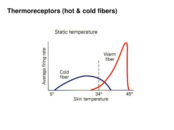
Drugs can mimic sensory stimuli Response of taste nerve to cold water or menthol
Pain • Pain and nociception • Pain - feeling of sore, aching, throbbing • Nociception - sensory process, provides signals that trigger pain • Nociceptors: Transduction of Pain • Bradykinin • Mast cell activation: Release of histamine
Nociception and the Transduction of Painful Stimuli • Types of Nociceptors • Polymodal nocireceptors, Mechanical nocireceptors, Thermal nocireceptors • Hyperalgeia • Primary and secondary hyperalgesia • Bradykinin, prostaglandins, and substance P
Primary Afferents and Spinal mechanisms First pain and second pain
Formalin paw-licking test Acute and tonic phases
Central Sensitization at first dorsal horn synapse
Central Sensitization at first dorsal horn synapse NMDA blockers Ibuprofen etc
Tonic Phase of formalin paw-licking requires spinal NMDA glutamate receptors
Ascending Pain Pathways • Differences between touch and pain pathway • Nerve endings in the skin • Diameter of axons • Connections in spinal cord • Touch – Ascends Ipsilaterally (crosses over in brainstem) • Pain – Ascends Contralaterally (crosses over right after entering spinal cord)
Ascending Pain Pathways (Cont’d) • Spinothalamic Pain Pathway • The Trigeminal Pain Pathway • The Thalamus and the Cortex • Touch and pain systems remain segregated • Pain and temperature information sent to various cortical areas
The Regulation of Pain • Afferent Regulation • Descending Regulation • The endogenuos opiates • Opioids and endomorphins
The Regulation of Pain (Cont’d) Descending regulation Opiates
Analgesia is the relief of pain. Pain is normally defined as an unpleasant sensory and emotional experience associated with potential or actual tissue damage. Pain is difficult to assess in animals because of the inability to communicate directly about what the animal is experiencing. Instead, indirect signs of pain are often used. Because of the difficulty of determining when an animal is in pain, animal welfare regulations require that analgesia be provided whenever a procedure is being performed or a condition is present that is likely to cause pain. In the absence of evidence to the contrary, it is assumed that something that is painful in a human will also be painful in an animal.
Assessment of pain or distress may be based on many different criteria including: • Decreased activity • Abnormal postures, hunched back, muscle flaccidity or rigidity • Poor grooming • Decreased food or water consumption • Decreased fecal or urine output • Weight loss (generally 20-25% of baseline), failure to grow, or loss of body condition (cachexia) • Dehydration • Decrease or increase in body temperature • Decrease or increase in pulse or respiratory rate • Physical response to touch (withdrawal, lameness, abnormal aggression, vocalizing, abdominal splinting, increase in pulse or respiration) • Teeth grinding (seen in rabbits and farm animals) • Self-aggression • Inflammation • Photophobia • Vomiting or diarrhea • Objective criteria of organ failure demonstrated by hematological or blood chemistry values, imaging, biopsy, or gross dysfunction
Anesthesia is a state of unconsciousness induced in an animal. The three components of anesthesia are analgesia (pain relief), amnesia (loss of memory) and immobilization. Curariform skeletal muscle relaxants or neuromuscular blockers (e.g. succinylcholine, decamethonium, curare, gallamine, pancuronium) are not anesthetics and have no analgesic effects. They may only be used in conjunction with general anesthetics. Normally, artificial respiration must be provided. Physiologic monitoring methods must also be used to assess anesthetic depth, as normal reflex methods will not be reliable.
Local Anesthetics: Block nerve impulses at the site of application, eg. lidocaine which blocks voltage-sensitive sodium channels Inhalation Anesthetics: Depress cortical activity by interactions with membranes of neurons Sedative Anesthetics: Provide sedation or anticonvulsant action by enhancing GABAergic chloride channels which inhibit cortical function, e.g. barbituates and benzodiazepines Dissociative Anesthetics: The dissociative anesthetics include ketamine (Vetalar, Ketaset) and tiletamine (Telazol). These drugs are easy to use and have a wide margin of safety for most laboratory species. They are cyclohexamine compounds, chemically related to piperazine and phencyclidine (PCP). The dissociative anesthetics uncouple sensory, motor, integrative, memory and emotional activities in the brain, providing there is a functional cerebral cortex. The state induced by high doses of ketamine is best described as catalepsy and is not accompanied by central nervous system depression. There is depression of respiratory function, but cardiovascular function is maintained. Muscle relaxation is very poor.
Muscle Afferents & Spinal Reflexes input -> processing -> output receptors -> afferents -> spinal cord -> efferents -> muscles afferents -> dorsal horn -> ventral horn -> efferents afferents = towards the spinal cord/brain efferents = away from the spinal cord/brain
flexor extensor
Muscle Afferents help control the action of muscles relay position of limbs (proprioception) convey sense of movement (kinesthesia) Two receptor organs: Spindles: stretch info via Ia and II afferents Golgi Tendon Organs: force info via IIb afferents
Muscle Spindle sensory organ in parallel with muscle fibers to provide info on muscle stretch (= length changes). Very dense in fine muscles (e.g. thumb). Highest density in neck, because head positition important. Fusiform bundle of 5-10 intrafusal muscle fibers (extrafusal = rest of muscle; fusus = spindle). Afferents = 1a sensory axons Efferents = alpha motor neurons to muscle = gamma motor neurons to spindle
Golgi Tendon Organ sensory organ in series with muscle fibers to provide info on muscle force (= load on the muscle). Located in tendons where muscle inserts on bone. Sensitive to changes in muscle force, but only to tension of a small number of motor units attached near to the tendon. Afferents = 1b sensory axons
Afferent Nerves cell bodies in dorsal root ganglion, enter dorsal horn of spinal cord. Ia afferent (primary afferent of spindle) large diameter, so high speed ( 12-20 µm, 72-120 m/sec). dynamic - responds to changes in length, peak firing rate proportional to velocity of stretch. Ib afferent large diameter that wraps around Golgi tendon organ. Responds to force. II afferent (secondary afferent of spindle) smaller diameters (4-12 µm, 24-72 m/sec). static –firing rate increases with increased stretch.
Alpha & Gamma Fibers alpha motor neurons: cause motor units to contract for skeletal muscle movements gamma motor neurons: cause intrafusal fibers to contract to maintain tension as fiber is stretched
Afferent Firing Patterns • Stretch -> increased firing of Ia & II afferents • Contraction • -> unloading of spindle • -> decreased Ia & II firing • -> increased tension on tendon, Golgi tendon organ • -> increased Ib firing
Spinal Reflexes Reflexes – stereotyped motor behaviors evoked by specific sensory stimuli • threshold of stimulus • applied to specific regions, motor response is stereotyped (not very variable) • only a few synapses, so not much processing and very fast • often under descending inhibition so revealed or exaggerated after spinal or brain injury (in dogs by Sherrington, after WWI by Head). • mediated by spinal cord or brainstem
Stretch Reflex (myotactic) skeletal muscle contracts, if stretched phasic component brief and quick response – e.g. knee jerk tonic component sustained over many seconds – hard to elicit in normal person


