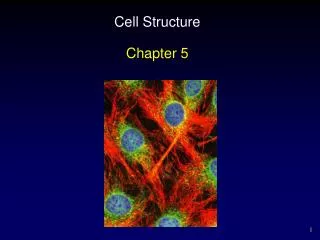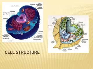
Cell Structure
Cell Structure. Melanie Pd. 8. Cells are the building blocks of all plants and animals. Cells are produced by the division of preexisting cells. Cells are the smallest units that perform all vital physiological functions. Each cell maintains homeostasis at the cellular level. Cell Theory.

Cell Structure
E N D
Presentation Transcript
Cell Structure Melanie Pd. 8
Cells are the building blocks of all plants and animals. • Cells are produced by the division of preexisting cells. • Cells are the smallest units that perform all vital physiological functions. • Each cell maintains homeostasis at the cellular level. Cell Theory
Outside the cell Within the cell Cytoplasm Cytosol Organelles • Extracellular fluid • What the cell floats in. Fluids
The cell membrane forms the outer boundary of the cell. • The most important components of the cell membrane include: • Phospholipids • Proteins • Glycolipids • Cholesterol. • The cell membrane is also called the plasma membrane, or sometimes even plasmalemma. Cell Membrane
All parts of the cell must work together to maintain homeostasis at the tissue, organ, and system levels. • All communication and coordination activities involve the cell membrane, because it forms the interface between each cell and its surroundings. • The cell membrane regulates the dynamic exchange between the intracellular and extracellular fluids. It is very important to maintain regulation between the two fluids, because each is different from the other, and in order to preserve homeostasis. A cell’s interaction
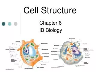
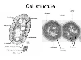
Cell Membrane- Lipid bilayer, containing phospholipids, steroids, and proteins. Isolation, protection, sensitivity, organization. • Cytosol- Fluid component of cytoplasm. Distributes materials by diffusion. • Cytoskeleton (microtubules, microfilaments)- Proteins organized in fine filaments or slender tubes. Strength, movement of cellular structures and materials. • Microvilli- Membrane extensions containing microfilaments. Absorption of extracellular materials. • Cilla- Membrane extensions containing microtubules in 9 X 2 arrangement. Movement of materials over surface. • Centrioles- Two centrioles, at right angles; each composed of microtubules in 9 X 3 array. Movement of chromosomes during cell division. • Ribosomes- RNA + proteins; fixed ribosomes bound to endoplasmic reticulum, free ribosomes scattered in cytoplasm.Protein synthesis. • Mitochondria- Double membrane, with inner folds (cristae) enclosing important metabolic enzymes. Produce 95% of the ATP required by the cell. Structure and functions of Organelles.
Nucleus- Nucleoplasm containing nucleotides, enzymes, and nucleoproteins; surrounded by double membrane or “nuclear envelope” • Nucleolus- Dense region in nucleoplasm. Site of RNA synthesis. • Endoplasmic reticulum- Network of membranous channels extending throughout the cytoplasm. Synthesis of secretory products; intracellular storage and transport. • Rough ER- Ribosomes attached to membranes. Secretory protein synthesis. • Smooth ER- Lacks attached ribosomes. Lipid and carbohydrate synthesis. • Golgi apparatus- Series of stacked, flattened membranes (saccules) containing chambers (cisternae). Storage, alteration, and packaging of secretory products and lysosomes. • Peroxisomes- Vesicles containing degradative enzymes. Neutralization of toxic compounds. Structure and functions of Organelles
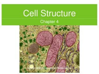
Cells obtain 95% of their necessary energy from a Membranous Organelle inside the cell named, Mitochondria. • The Mitochondria is composed of Double Membrane, with inner folds (cristae) enclosing important metabolic enzymes. • The Mitochondria produces 95% of the ATP required by the cell. Energy of a Cell.
The Nucleus is composed of Nucleoplasm containing nucleotides, enzymes, and nucleoproteins; surrounded by double membrane or “nuclear envelope”. • It controls the metabolism of the cell. • It also controls storage and processing of genetic information. Nucleus.
The cell-division cycle is a essential process where a single-celled organism splits to make another organism. • In cells with a nucleus, the cycle is divided into two periods: • Interphase: • The cell grows, accumulates nutrients needed for mitosis and duplication its DNA. • Mitosis (M) Phase: • The cell splits itself in two, which are often called “daughter cells” • Cytokinesis- where the cell is completely divided. Cell Cycle.
Many cell functions that involve the cell membrane, such as secretion or ciliary movement, involve changes in the transmembrane potential. • Also, because the transmembrane potential can magnify a stimulus in this way, it greatly increases the cell’s sensitivity to its environment. Transmembrane Potential.
Two factors, one passive and one active, maintain the transmembrane potential. • The passive factor- is that the membrane permaebilties for sodium and potassium are quite different, thus causing the cell to lose positive charge faster than it can gain them. • Because these molecules are too large to pass through the membrane, the interior of the cell develops an excess of negative charges. • The active factor- is the presence of the sodium potassium exchange pump in the membrane surface. Again the potassium ions diffuse out of the cell much faster than sodium ions enter it, and the exchange pump cannot prevent the net loss of positive charges. • But because the rate is precisely balanced by the activity of the sodium-potassium exchange pump, the transmembrane potential is stabilized at this value. Maintaining the Transmembrane Potential
Most cells in the body attach to other cells or to extracellular protein fibers. • The attachment occurs at cell junctions, that are not involved in membrane flow. • There are four different types of cell junctions: • Gap Junction • Tight Junctions • Intermediate Junctions • Desmosomes Cell Attachment.
In Gap junction, two cells are held together by an interlocking of membrane proteins. • Because these are channel proteins, the result is a narrow passageway that lets small molecules and ions pass from cell to cell. • Gap Junctions are most common in: • Cardiac muscle • Smooth muscle tissue. • Occasionally found between nerve cells. Gap Junction:
At tight junctions there is a partial fusion of the lipid portions of the two cell membranes. • Because the membranes are fused together, tight junctions are the strongest intercellular connections. • Tight junctions also provide mechanical strength, block the passages of water or solutes between the cells. • They are often found: • Where cells are exposed to fluids whose composition is very different from that of normal extracellular fluid. Tight Junctions:
At intermediate junctions the opposite cell membrane are held together by a thick layer of proteoglycans. • This Proteoglycan layer is called, intercellular cement. • Hyaluronic acid is the most important proteoglycan involved. • The cytoplasm at one of these junctions contains a dense network of microfilaments that anchor the junction to the cytoskeleton. This arrangement adds strength and helps stabilize the shape of the cell. Intermediate Junctions:
At desmosomes these is a very thin proteoglycan layer between the opposing cell membranes, reinforced by a network of intermediate filaments that lock the two cells together. • Desmosomes are very strong and the connection can resist stretching and twisting. • These intercellular connections are most abundant between the cells in the superficial layers of the skin. Desmosomes:
Cytology- the study of the structure and function of cells. • Transmission electron microscopy- when electrons pass through an ultrathin section to strike a photographic plate. • Scanning electron microscopy- when electrons bouncing off exposed surfaces create a scanning electron micrograph. Vocabulary:
Extracellular fluid- a watery medium our cells float in. • Cell membrane or Plasma membrane- the outer boundary of the cell. • Cell membrane is also called a phospholipid bilayer, because they form two layers. • Peripheral proteins- attached to the inner membrane surface. • Integral proteins are embedded in the membrane. • Some of these proteins form channels that let water molecules, small water-solution compounds, and ions into and out of the cell.
Cytosol or intracellular fluid- contains dissolved nutrients, ions, soluble and soluble proteins and waste products. • The membrane also separates the cytosol from the surrounding extracellular fluid. • Organelles- structures that preform specific functions within the cell. • Inclusions- masses of insoluble materials sometimes found in the cytosol. • Nonmembranous organelles- cellular organelles that are always in contact with the cytosol. • Membranous organelles- cellular organelles that are surrounded by lipid membranes that isolate them from the cytosol.
Cytoskeleton- is an internal protein framework that gives the cytoplasm strength and flexibility. • Microfilaments- slender protein strands, usually composed of the protein actin. • Myosin- protein component of the thick myofilaments. • Neurofilaments- found in nerve cells, where they provide structural support and provably assist in the movement of materials within the cytoplasm. • Thick filaments- are relatively massive strands composed of myosin protein subunits. • Microtubules- found in all our cells are hollow tubes built from the globular protein tubulin. • Microvilli- small, finger-shaped projections of the cell membrane.
Centriole- is a cylindrical structure composed of short microtubules. • Centrosome- is the cytoplasm surrounding the pair. • Cilia- contains nine parts of microtubules surrounding a central pair. • Basal body- a compact situated just beneath the cell surface which the cilla are anchored to. • Flagella- resemble cilla but are much larger. • Ribosomes- are small, dense structure that cannot be seen clearly with the light microscope. • Free ribosomes- scattered throughout the cytoplasm, they product proteins that enter the cytosol. • Fixed ribosomes- are attached to the endoplasmic reticulum , a membranous organelle.
Mitochondria- small organelles that have an unusual double membrane. • Cristae- numerous folds in the inner membrane. • Matrix-the fluid contents of the mitochondrion. • Respiratory enzymes-attached to the cristae produce most of the ATP generated by mitochondria. • Nucleus- the control center for cellular operations. • Nuclear envelope- surrounds the nucleus and separates it from the cytosol. • Peri-nuclear space- a narrow passage in the double membrane in the nuclear envelope. • Nuclear pores- chemical communication between the nucleus and cytosol occurs here. • Nucleoplasm- the fluid contents of the nucleus.
Chromosomes- complex structures formed by the DNA. • Histones- chromosomes that contain DNA strands bound to special proteins. • Nucleosome-DNA strands wind around the histones at intervals, forming a complex known as Nucleosome. • Chromatin- in cells that are not dividing, when chromosomes are loosely coiled, forming a tangle of fine filaments. • Nucleoli- one to four dark-staining areas found in the nucleoli. • Endoplasmic reticulum- a network of intracellular membranes. • Cisternae- round chambers formed by the ER. • Rough endoplasmic reticulum- used as a combination workshop and shopping depot.