Effects of PC and APC on RASF Viability Following siRNA Transfection and Treatment
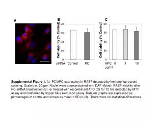
This study investigates the expression of PC/APC in rheumatoid arthritis synovial fibroblasts (RASF) using immunofluorescent staining. Nuclei are counterstained with DAPI (blue) to visualize cellular structure. Viability post siRNA transfection and recombinant APC treatment is assessed by MTT assay and confirmed with trypan blue exclusion. Results are presented as percentage of control, indicating no significant statistical differences observed. These findings provide insight into the role of PC and APC in RASF viability and potential therapeutic avenues.

Effects of PC and APC on RASF Viability Following siRNA Transfection and Treatment
E N D
Presentation Transcript
B C A Cell viability (% Control) Cell viability (% Control) siRNA Control PC APC 0 1 10 µg/ml Supplemental Figure 1. A) PC/APC expression in RASF detected by immunofluorescent staining. Scale bar: 20 µm. Nuclei were counterstained with DAPI (blue). RASF viability after PC siRNA transfection (B) or treated with recombinant APC (C) for 72 hrs detected by MTT assay and confirmed by trypan blue exclusion assay. Data on graphs are expressed as percentages of control and shown as mean ± SD (n=3). There were no statistical differences.