Download
1 / 1
Télécharger la présentation 
Specific Recognition of MUC20 Overexpression by anti-MUC20 Antibody
An Image/Link below is provided (as is) to download presentation
Download Policy: Content on the Website is provided to you AS IS for your information and personal use and may not be sold / licensed / shared on other websites without getting consent from its author.
Content is provided to you AS IS for your information and personal use only.
Download presentation by click this link.
While downloading, if for some reason you are not able to download a presentation, the publisher may have deleted the file from their server.
During download, if you can't get a presentation, the file might be deleted by the publisher.
E N D
Presentation Transcript
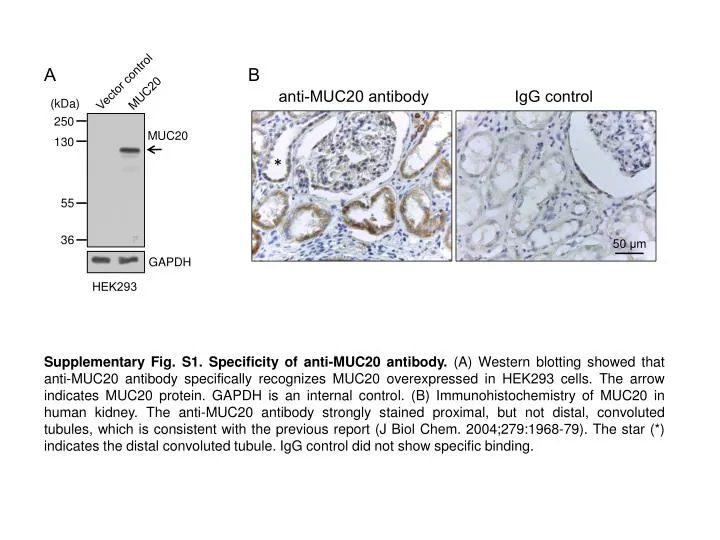
A B Vector control MUC20 anti-MUC20 antibody IgG control (kDa) 250 MUC20 130 * 55 36 50 μm GAPDH HEK293 Supplementary Fig. S1. Specificity of anti-MUC20 antibody. (A) Western blotting showed that anti-MUC20 antibody specifically recognizes MUC20 overexpressed in HEK293 cells. The arrow indicates MUC20 protein. GAPDH is an internal control. (B) Immunohistochemistry of MUC20 in human kidney. The anti-MUC20 antibody strongly stained proximal, but not distal, convoluted tubules, which is consistent with the previous report (J Biol Chem. 2004;279:1968-79). The star (*) indicates the distal convoluted tubule. IgG control did not show specific binding.


More Related