Download
1 / 33
330 likes | 471 Vues
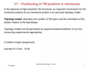
V7 – Positioning of TM proteins in membrane. In the absence of high-resolution 3D structures, an important cornerstone for the functional analysis of any membrane protein is an accurate topology model.

E N D
V7 – Positioning of TM proteins in membrane In the absence of high-resolution 3D structures, an important cornerstone for the functional analysis of any membrane protein is an accurate topology model. Topology model: describes the number of TM spans and the orientation of the protein relative to the lipid bilayer. Topology models can be generated by sequence-based prediction or by time-consuming experimental approaches. 2 medium length assignments tutorials Fri 13.00 - 14.30 Membrane Bioinformatics – Part II
(1) Global Topology Analysis Idea: generate reference point, e.g. the location of a protein‘s C terminus. In E.coli attach alkaline phosphatase (PhoA) that is active only in the periplasm of E.coli, or green fluorescent protein (GFP) that fluoresces only in the cytoplasm. TMHMM: 1000 of 4288 predicted E.coli genes are inner membrane proteins. 737 genes encode proteins with > 100 residues and 2 TM helices. 714 were suitable for cloning into phoA and gfp fusion vectors. Both fusions could be obtained for 573 genes, one fusion for an additional 92 genes. Daley et al. Science 308, 1321 (2005) Membrane Bioinformatics – Part II
Global Topology Analysis Using homology, 601 proteins could be assigned a topology. For 71 of these, the location of the C terminus was already established. The results agreed except for 2 cases. The error rate is therefore ~ 1%. TMHMM alone predicts the correct C-terminal location for 78% of the 601 proteins. By providing unambiguous C-terminal locations, the TMHMM reliability score increases for 526 proteins and decreases for 75 proteins. Daley et al. Science 308, 1321 (2005) Membrane Bioinformatics – Part II
Functional categorization of E.coli inner membrane proteome clear trend for Nin – Cin topologies (even number of TMH) - largest functional category is transport proteins, many with 6 or 12 TM helices. Most proteins with unknown function have 6 TM helices. Daley et al. Science 308, 1321 (2005) Membrane Bioinformatics – Part II
Extend predictions by sequence homology Idea: transfer experimental data set from PhoA and GFP-fusions to homologous proteins. Data on 608 proteins. 204 annotated eubacterial and 21 archeal genomes in March 2005, 658,210 sequences. BLAST searches (E-value < 10-5) 30,744 sequence hits where TMHMM predicts 1 TM helix Second BLAST query with these 30,744 sequences 17,111 „secondary homologs“. Granseth et al., J.Mol.Biol. 352, 489 (2005) Membrane Bioinformatics – Part II
Unconstrained vs. constrained prediction (a) Unconstrained TMHMM predictions for the full set of 158,182 sequences with 1 predicted TM helix (grey bars) and constrained predictions for the 51,208 sequences for which the C-terminal location or the location of an internal residue could be annotated (black bars). The number of proteins with different topologies are shown; Cin topologies are plotted upwards, Cout downwards. The number of Cout proteins with a single TM helix (39,322) is off-scale. The unconstrained algorithm predicts too many proteins as Cout. (b) TMHMM predictions for the 51,208 annotated sequences before (grey bars) and after (black bars) constraining the predictions with the location of the annotated residue. Granseth et al., J.Mol.Biol. 352, 489 (2005) Membrane Bioinformatics – Part II
(2) Dual-topology proteins? Most TM proteins are expected to adopt only one topology in the membrane. Global topology analysis of E.coli inner membrane proteome identified 5 dual-topology candidates: EmrE, SugE, CrcB, YdgC, YnfY. All are quite small (~ 100 aa), contain 4 strongly predicted TM segments, contain only few K and R residues and have very small (K + R) bias. (a) A dual-topology protein inserts into the membrane in two opposite directions. As nearly all helix-bundle membrane proteins have a higher number of lysine (K) and arginine (R) residues in cytoplasmic (in) than in periplasmic (out) loops (the ‚positive-inside‘ rule), dual-topology proteins are expected to have very small (K + R) biases. Rectangles: TM segments black dots: K and R residues Rapp et al., Nat.Struct.Biol. 13, 112 (2006) Membrane Bioinformatics – Part II
Dual-topology proteins? Without solving their 3D structures, how can one prove that a protein has dual topology? Such a protein would be particularly sensitive to the addition or removal of a single positively charged residue in a loop or tail. measure activities of two different, C-terminally fused reporter proteins: PhoA (only enzymatically active when in the periplasm) GFP (fluorescent only when in the cytoplasm). Concentrate on N-terminus and first loop. Rapp et al., Nat.Struct.Biol. 13, 112 (2006) Membrane Bioinformatics – Part II
Charge mutations shift the orientations of dual-topology TM proteins (a) wt YdgE-PhoA fusion is active, wt YdgE-GFP fusion is inactive C-terminus in periplasm (Cout ) wt YdgF behaves oppositely (Cin) These 2 proteins are topologically stable. (b – d) C-terminal orientation of EmrE, SugE, CrcB, YnfA and YdgC is highly sensitve to charge mutations. For 14 or 19 charge mutations, both PhoA and GFP activities change in the direction expected from the change in (K + R) bias. Rapp et al., Nat.Struct.Biol. 13, 112 (2006) Membrane Bioinformatics – Part II
Dual-topology homologs occur as gene pairs or singletons Pfam searches in 174 fully sequenced bacterial genomes for homologs (E < 10-10) to SugE, EmrE, YdgE, CrcB, YnfA, YdgC and YdgO/YdgL. Create multiple sequence alignment with ClustalW. Use TMHMM to predict the positions of TM helices. Obtain consensus TM helix prediction, compute (K + R) biases for individual proteins. 10 residues from each of the flanking TM helices were included to allow for possible misprediction of the exact positions of the loop ends. Rapp et al., Nat.Struct.Biol. 13, 112 (2006) Membrane Bioinformatics – Part II
Dual-topology homologs occur as gene pairs or singletons Interpretation: SMR and CrcB occur as closely spaced pairs or as singletons. Paired genes encode homologous proteins with opposite (K + R) bias. Rapp et al., Nat.Struct.Biol. 13, 112 (2006) Membrane Bioinformatics – Part II
An internally duplicated protein with opposite topology Most likely evolutionary scenario: a single dual-topology protein undergoes gene duplication, the two resulting proteins become fixed in opposite orientations and finally fuse into a single polypeptide. Rapp et al., Nat.Struct.Biol. 13, 112 (2006) Membrane Bioinformatics – Part II
(3) Prediction of buried TM helices Global topology analysis of E.coli inner membrane proteome showed that ca. 20 – 25% of the TM proteins have 10 TM helices. These are often involved in transport of small molecules across the membrane. Many of these proteins will have buried helices. Can we identify those? Develop an empirical helix burial function f based on a few assumptions. (i) residues in buried helices are more conserved because of structural and functional contraints. (ii) the residue composition of the buried helices is different from the composition of helices facing the lipid environment. (iii) the difference between the minimal and maximal values of conservation entropy for every position in MSAs of TM helices should be smaller in buried helices than in lipid-exposed helices because of the homogenous environment. Adamian & Liang, Proteins 63, 1 (2006) Membrane Bioinformatics – Part II
Burial Function f: burial function s: average entropy of all residue positions of the TM helix l : average lipophilicity k: sorted entropy values of all residue positions in a helix of length d for helices 1 ... n of the TM protein Problems: the average entropy depends on the number of sequences in the MSA. needs MSAs with exactly the same set of sequences from the same set of species. Also, the stability of different membrane proteins in the lipid environment may be different. Account for ambiguity in the definition of TM helix ends. Adamian & Liang, Proteins 63, 1 (2006) Membrane Bioinformatics – Part II
Ranking of TM helices by burial function and robustness Adamian & Liang, Proteins 63, 1 (2006) Membrane Bioinformatics – Part II
Examples of buried TM helices that are correctly predicted (a) TM helices TM4, TM5, TM6, TM8 form core, consistent with prediction. (b) TM4, TM10 are most buried. (c) one can explain prediction of TM8 as buried by considering a tightly bound cardiolipin molecule identified in the X-ray structure. Adamian & Liang, Proteins 63, 1 (2006) Membrane Bioinformatics – Part II
Test ranking results Is the method applicable to TM proteins where only sequence data is available? Test on structure of Leu transporter. TMHMM predicts 12 TM helices. Good overlap with X-ray helices. Problem that no additional sequences exist that are annotated as Na+-dependent Leu transporters. LeuTAa has 3 significantly buried helices: 1, 6 and 8. 1 and 6 are true positives, 2 is a false positive, 8 is a false negative. Adamian & Liang, Proteins 63, 1 (2006) Membrane Bioinformatics – Part II
(4) Positioning of proteins in membranes Experimental techniques to study orientation of proteins in membranes chemical modification spin-labeling fluorescence quenching X-ray scattering neutron diffraction electron cryomicroscopy NMR polarized infrared spectroscopy. Desirable to complement by computational methods. e.g. explicit-solvent molecular dynamics ... up to simplified approaches that minimize the protein transfer energy from water to a hydrophobic slab (used as a membrane model). Adamian & Liang, Proteins 63, 1 (2006) Membrane Bioinformatics – Part II
important parameters Lomize et al. Prot.Sci. 15, 1318 (2006) Membrane Bioinformatics – Part II
Calculation of transfer energy Model protein as a rigid body that freely floats in the planar hydrocarbon core of a lipid bilayer. ASAi : accessible surface area of atom i, computed with NACCESS iW-M : solvation parameter of atom i (transfer energy of the atom from water to membrane interior in kcal/(mol.Å2) ) f(zi): interfacial water concentration profile with = 0.9 Å Adamian & Liang, Proteins 63, 1 (2006) Membrane Bioinformatics – Part II
ionization of charged residues Residues that are typically charged in soluble proteins may become neutral in the hydrophobic inside of the bilayer! The ionization/protonation energies of charged residues are described by the Henderson-Hasselbalch equation: at pH = 7 average pKa value Gioniz in proteins [kcal/mol] Arg 12.0 6.9 Lys 10.4 4.7 Asp 3.4 4.9 Glu 4.1 4.0 His 6.6 0.6 Lomize et al. Prot.Sci. 15, 1318 (2006) Membrane Bioinformatics – Part II
Global energy optimization use deterministic 2-step search strategy: (1) grid scan to determine a set of low-energy combinations of variables z0, d, , grid steps: 0.5 Å for z0 and d, 5° for , 2° for (2) local energy minimization (Davidon-Fletcher-Powell method) starting from low-energy points Also consider energetically best rotation of solvent-exposed charged side chains (e.g. Lys and Arg) that are situated close to the calculated boundaries and could be rotated away from the hydrophobic core Adamian & Liang, Proteins 63, 1 (2006) Membrane Bioinformatics – Part II
Which solvation parameters to use? chx and dcd results agree well with experiment, oct agrees poorly. Lomize et al. Prot.Sci. 15, 1318 (2006) Membrane Bioinformatics – Part II
features of model slightly different parameter sets should be applied for proteins in detergents and bilayers Gtransfer should not include contributions of atoms that face internal polar cavities of TM proteins and that do not directly interact with surrounding bulk lipid ( mention results of Sam) Otherwise, the orientation of many -barrels and pore-forming transporters would be computed incorrectly Lomize et al. Prot.Sci. 15, 1318 (2006) Membrane Bioinformatics – Part II
Main features of model necessary and sufficient approximations for reproducing the exp. data (1) lipid bilayer is represented as planar hydrophobic slab with adjustable thickness and a narrow interfacial area with a sigmoidal polarity profile (2) proteins are considered as rigid bodies with flexible side chains; their transfer energies are minimized with respect to 4 variables (3) transfer free energy is calculated at an all-atom level using atomic solvation parameters determined for the water-decadiene system (4) neglect explicit electrostatic interactions, account for neutralization of charged residues (5) eliminate contributions of pore-facing atoms The model only depends on 5 atomic solvation parameters (N, O, S, sp2 C, sp3 C), one constant , and the ionization energies of charged groups. All can be obtained independently from experimental sources. Verify method for 24 TM proteins of known 3D structure whose spatial position in bilayers have been exp studied. Lomize et al. Prot.Sci. 15, 1318 (2006) Membrane Bioinformatics – Part II
Average tilt angles (a) hydrophobic thickness matches well (table 2) (b) the calculated tilt values are in excellent agreement with NMR data, they also correlate well with ATR-FTIR data (table 3), although the exp. values are systematically larger orientational disorder in the experiments? Lomize et al. Prot.Sci. 15, 1318 (2006) Membrane Bioinformatics – Part II
Membrane penetration depths Lomize et al. Prot.Sci. 15, 1318 (2006) Membrane Bioinformatics – Part II
Introduction Lomize et al. Prot.Sci. 15, 1318 (2006) Membrane Bioinformatics – Part II
Membrane core boundaries Lomize et al. Prot.Sci. 15, 1318 (2006) Membrane Bioinformatics – Part II
Application to all TM proteins from the PDB application to all other 109 TM protein complexes 80 -helical 28 -barrels gramicidin dimer control set: 20 water-soluble proteins 32 monotopic and peripheral proteins Lomize et al. Prot.Sci. 15, 1318 (2006) Membrane Bioinformatics – Part II
Application to membrane proteins Peripheral and monotopic proteins have low penetration depths. Calculated tilt angles vary from 0° - 6°. TM proteins tend to be nearly perpendicular to the membrane, although the individual helices are on average tilted by 21°. Lomize et al. Prot.Sci. 15, 1318 (2006) Membrane Bioinformatics – Part II
Biological membranes differ Lomize et al. Prot.Sci. 15, 1318 (2006) Membrane Bioinformatics – Part II
Fluctuations around energy minimum Fluctuations are larger for TM proteins with a smaller TM perimeter. Lomize et al. Prot.Sci. 15, 1318 (2006) Membrane Bioinformatics – Part II