Download
1 / 43
450 likes | 706 Vues
Learn about serous, fibrinous, suppurative inflammations, abscesses, ulcers, and pseudomembranous inflammation variations.

E N D
MORPHOLOGIC PATTERNS OF ACUTE INFLAMMATION DR.AYSER HAMEED LEC.3
Many variables may modify the basic inflammatory response; these include:- 1. The nature and intensity of the injury. 2. The site and tissues affected. 3. The responsiveness of the host. Several types of inflammation are recognized, which vary in their morphology and clinical correlates.
Serous inflammation Is characterized by the outpouring of a thin fluid that is derived from either the plasma or the secretions of mesothelial cells lining the peritoneal, pleural, and pericardial cavities. In these serous cavities the accumulated fluid is called effusion. The skin blister resulting from a burn or viral infection represents a large accumulation of serous fluid, either within or immediately beneath the epidermis of the skin.
Serous inflammation (serous pleural effusion) Excessive accumulation of clear, thin fluid within pleural cavity. It is transparent but note the reflection of light in the upper part of the photograph and lung collapse (arrow) due to pressure induced by the fluid.
Fibrinous inflammation With more severe injuries and the resulting greater vascular permeability, larger molecules such as fibrinogen pass the vascular barrier, and fibrin is formed and deposited in the extracellular space. A fibrinousexudate develops in such cases. The latter also occurs when there is a stimulus for coagulation in the interstitium (e.g. cancer cells). A fibrinousexudate is characteristic of inflammation in the lining of body cavities, such as the meninges, pericardium, and pleura.
Fibrinousexudate-pericardium • there is a lot of fibrin. • the visceral and parietal surfaces become stuck together (by fibrin). • Separation of the two layers imparts rough irregular appearance (the so called bread and butter).
Microscopically:fibrin appears as an eosinophilic meshwork of threads or amorphous coagulated mass. Fibrinous exudates may be removed by fibrinolysis and clearing of other debris by macrophages. However, when the fibrin is not removed, it may stimulate the ingrowth of fibroblasts and blood vessels and thus lead to scarring.
Conversion of the fibrinousexudate to scar tissue is called organization. When this occurs within the pericardial sac it leads either to opaque fibrous thickening of the pericardium or, more often, to the development of fibrous strands that reduce and may even obliterate the pericardial space.
Suppurative (purulent) inflammation This is characterized by the production of large amounts of pus or purulent exudate consisting of neutrophils, necrotic cells, and edema fluid. Certain bacteria (e.g., staph. aureus, St. pyogenes, Pneumococci, gonococci, meningococci and E. coli) produce this localized suppuration and are therefore called pyogenic (pus-producing) bacteria. A common example of an acute suppurative inflammation is acute (suppurative) appendicitis.
Appendix: acute suppurative inflammation Upper half of excised appendix. Lt: fibrino-purulent serosalexudateRt: lumen filled with pus
An abscess is a localized collection of purulent inflammatory fluid (pus) caused by suppuration buried in a tissue, an organ, or a confined space. Pus is a thick creamy yellow or blood-stained fluid. Abscesses are produced by deep seeding of pyogenic bacteria into a tissue. They have a central region that appears as a mass of necrotic leukocytes and tissue cells.
There is usually a zone of preserved neutrophils around this necrotic focus, and outside this region vascular dilation and fibroblastic proliferation occur, indicating the beginning of repair. In time, the abscess may become walled off and ultimately replaced by connective tissue. A common example of an abscess is the skin furuncle.
Abscess (Furuncle) (boil) Abscess that involves the skin is called “Boil” or “furuncle”.
Ulcers :- An ulcer is a local defect, or excavation of the surface of an organ or tissue that is produced by the sloughing (shedding) of inflammatory necrotic tissue. Ulceration occurs only when tissue necrosis and resultant inflammation exist on or near a surface.
It is most commonly encountered in: 1. Inflammatory necrosis of mucosa-lined cavities e.g. mouth, larynx, stomach, intestines, or genitourinary tract. 2. Subcutaneous inflammation of the lower extremities in older persons who have circulatory disturbances that predispose to extensive necrosis. Ulcerations are best exemplified by peptic ulcer of the stomach or duodenum, in which acute and chronic inflammation coexist.
Chronic peptic ulcer stomach Sharply delimited chronic peptic ulcer with converging folds of mucosa in the upper half
Pseudomembranous inflammation of mucous membranes. Severe injury may be associated with extensive epithelial necrosis with sloughing. This creates large shallow ulcers. Fibrin, dead epithelium, neutrophils, red cells and bacteria mix together to produce a white or cream-colored false (pseudo-) membrane covering the affected mucosa. Diphtheria and psudomembranous colitis are typical examples.
Pseudomembranousentercolitis ulcerations pseudomembrane This yellow-green exudate on the surface of an inflamed, hyperemic (erythematous) bowel mucosa consists of many neutrophils along with fibrin and amorphous debris from dying cells.
EFFECTS OF ACUTE INFLAMMATION Beneficial Effects:- 1. Dilution of Toxins by the edema fluid. 2. Production of protective Antibodies & promotion of immunity. 3. Fibrin meshwork formation that forms a scaffold for inflammatory cell migration & also limits the spread of infections. 4. Cell Nutrition.
Harmful Effects 1. Swelling & edema that can be detrimental for e.g. acute epiglottitis that may be life threatening. 2. Rise in tissue pressure that contributes to tissue necrosis. 3. Digestion of adjacent viable tissue. 4. Sever damaging allergic reaction. 5. Generalized increase in vascular permeability can cause shock as seen in anaphylactic reactions.
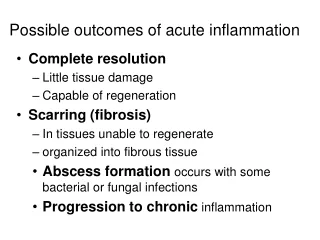
OUTCOMES OF ACUTE INFLAMMATION In general, acute inflammation may have one of three outcomes :- 1. Complete resolution. The battle between the injurious agent and the host may end with restoration of the site of acute inflammation to normal. This is called resolution and is the usual outcome when:- a. the injury is limited or short-lived. b. there has been little tissue destruction. c. the damaged parenchymal cells can regenerate.
2. Healing by fibrosis This occurs :- a. after extensive tissue destruction. b. when the inflammatory injury involves tissues that are incapable of regeneration. c. when there is abundant fibrin exudation. When the fibrinousexudate in tissue or serous cavities (pleural, peritoneal, synovial) cannot be adequately cleared, connective tissue grows into the area of exudate, converting it into a mass of fibrous tissue—a process also called organization.
3. Progression to chronic inflammation Acute to chronic transition occurs when the acute inflammatory response persists, owing either to the perseverance of the injurious agent or to some interference with the normal process of healing. For example, failure of acute bacterial pneumonia to resolve may lead to extensive tissue destruction and formation of a cavity in which the inflammation continues to smolder, leading eventually to a chronic lung abscess.
CHRONIC INFLAMMATION Although it may follow acute inflammation, it frequently begins from the outset as a chronic (chronic inflammation ab initio), insidious, and low-grade, smoldering response. Chronic inflammation is the cause of tissue damage in some of the most common and disabling human diseases, such as rheumatoid arthritis, atherosclerosis, tuberculosis, and chronic lung diseases.
Chronic Inflammation may complicate acute inflammation. The latter is almost always a suppurative type of inflammation that presents as a purulent discharge (pus) as seen in abscess. The cause is either a delay in the evacuation of an abscess, or presence of foreign-body within inflamed area (dirt, wood, metal or a sequestrated bone).
Causes of chronic inflammation ab initio include:- 1. Persistent infections by certain microorganismssuch as tubercle bacilli, Treponemapallidum, certain viruses, fungi, and parasites. These organisms are of low toxicity and evoke delayed type hypersensitivity reaction. 2. Prolonged exposure to toxic agents either exogenous as inhaled silica particles, or endogenous such as toxic plasma lipids that are thought to be responsible for atherosclerosis. The latter is thought to be a chronic inflammatory process of the arterial wall. 3. Autoimmunity.
Under certain conditions, immune reactions develop against the individual's own tissues, leading to autoimmune diseases. In these diseases, autoantigens activate a self-perpetuating immune reaction that results in chronic inflammation with associated tissue damage. Examples of this type include several common chronic inflammatory diseases, such as rheumatoid arthritis and lupus erythematosus. Morphologic features of chronic inflammation :- In contrast to acute inflammation, which is manifested by vascular changes, edema, and predominantly neutrophilic infiltration,
chronic inflammation is characterized by: 1. Infiltration with mononuclear cells including macrophages, lymphocytes, and plasma cells. 2. Tissue destruction, induced by the persistent offending agent or by the inflammatory cells. 3. Attempts at healing by fibrosis of the damaged tissue, achieved by proliferation of small blood vessels (angiogenesis) & fibroblasts.
Features of chronic inflammation The three characteristic features of chronic inflammation (in the lung) Chronic inflammatory cells infiltration.* Destruction of the normal tissue. (normal alveoli are replaced by spaces lined by cuboidal cells (arrow heads). 3. Replacement by fibrosis (arrows).
Mononuclear cell infiltration The macrophage is the dominant cells in chronic inflammation. The mononuclear phagocyte system (reticuloendothelial system) consists of closely related cells of bone marrow origin, including blood monocytes and tissue macrophages. The latter are diffusely scattered in connective tissues or located in organs such as the liver (Kupffer cells), spleen and lymph nodes (sinus histiocytes), and lungs (alveolar macrophages). From the blood, monocytes migrate into various tissues and differentiate into macrophages.
The half-life of blood monocytes is about 1 day, whereas the life span of tissue macrophages is several months or years. When the monocyte reaches the extravascular tissue, it undergoes transformation into a larger phagocytic cell, the macrophage. Macrophages may be activated by a variety of stimuli, including cytokines (e.g., IFN-γ) secreted by sensitized T lymphocytes, NK cells, bacterial endotoxins, and other chemical mediators.
Activation results in increased cell size, and greater ability to phagocytose and kill ingested microbes. Activated macrophages secrete a wide variety of biologically active products that result in the tissue injury and fibrosis. In short-lived acute inflammation, if the irritant is eliminated, macrophages eventually disappear (dying off or travel through lymphatics to lymph nodes). In chronic inflammation, macrophage accumulation persists, and this is mediated by the following:-
1. Recruitment from circulating monocytes; a process fundamentally similar to that of neutrophils. 2. Local proliferation of macrophages after their emigration from the bloodstream. This is now known to occur prominently in some chronic inflammatory lesions, such as atheromatous plaques. 3. Immobilization of macrophages within the site of inflammation. Certain cytokines and oxidized lipids can cause such immobilization (migration inhibiting factors).
The products of activated macrophages serve to eliminate injurious agents such as microbes and to initiate the process of repair, but are also responsible for much of the tissue injury in chronic inflammation; these products include:- 1. Toxic substances to microbes and host cells (e.g. toxic O2 species, NO, and proteases). 2. Chemoattractantsto other inflammatory cells. 3. Growth factors: the cause of fibroblast proliferation, collagen deposition, and angiogenesis.
Other cells in chronic inflammation :- Other cell types present in chronic inflammation include lymphocytes, plasma cells, eosinophils, and mast cells: Lymphocytes are mobilized in immune and nonimmune inflammation. Antigen-stimulated T and B-cells use various adhesion molecules (predominantly the integrins) and chemokines to migrate into inflammatory sites.
Lymphocytes and macrophages interact in a bidirectional way and these reactions play an important role in chronic inflammation. Macrophages display antigens to T cells that stimulate them. Activated T lymphocytes produce cytokines, and one of these, IFN-γ, which is a major activator of macrophages.
Plasma cellsdevelop from activated B lymphocytes and produce antibody directed against persistent antigen in the inflammatory site. Eosinophilsare abundant in immune reactions mediated by IgE and in parasitic infections. The recruitment of eosinophils involves extravasation from the blood and their migration into tissue by processes similar to those for other leukocytes. One of the chemokines that is especially important for eosinophil recruitment is eotaxin. Eosinophils have granules that contain major basic protein that is toxic to parasites.
Mast cellsare widely distributed in connective tissues and participate in both acute and persistent inflammatory reactions. Mast cells express on their surface the receptor that binds the Fc portion of IgE antibody. In acute reactions, IgE antibodies bound to the cells' Fc receptors specifically recognize antigen, and the cells degranulate and release mediators, such as histamine and products of AA oxidation. Mast cells are also present in chronic inflammatory reactions, and may produce cytokines that contribute to fibrosis.
Neutrophilsalthough characteristic of acute inflammation, many forms of chronic inflammation continue to show large numbers of neutrophils, induced either by persistent microbes or by mediators produced by macrophages and T lymphocytes. In chronic bacterial infection of bone (osteomyelitis), a neutrophilicexudate can persist for many months. Neutrophils are also important in the chronic damage induced in lungs by smoking and other irritant stimuli.
Examples of chronic inflammation Chronic Cholecystitismay be the sequel to repeated bouts of acute cholecystitis, but in most instances it develops de novo. Like acute cholecystitis it is almost always associated with gallstones but these do not seem to have a direct role in the initiation of inflammation. Rather, supersaturation of bile predisposes to both chronic inflammation and, in most instances, stone formation.
Microorganisms, usually E. coli and enterococci, can be cultured from the bile in only about one-third of cases. The gallbladder may be contracted, of normal size, or enlarged. The submucosa and subserosa are often thickened from fibrosis. In the absence of superimposed acute cholecystitis, mural lymphocytes are the only feature of inflammation.
Chronic cholecystitis with cholelithiasis Note thickening of the wall due to fibrosis