Download

1 / 37
370 likes | 395 Vues
Learn about using GFP in bacterial transformation & protein purification, its real-world uses, and electrophoresis procedures.

E N D
Instructors Stan Hitomi Coordinator – Math & Science Principal – Alamo School San Ramon Valley Unified School District Danville, CA Kirk Brown Lead Instructor, Edward Teller Education Center Science Chair, Tracy High School and Delta College, Tracy, CA Bio-Rad Curriculum and Training Specialists: Sherri Andrews, Ph.D. sherri_andrews@bio-rad.com Essy Levy, M.Sc. essy_levy@bio-rad.com Leigh Brown, M.A. leigh_brown@bio-rad.com
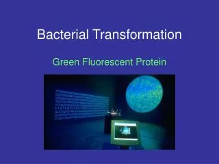
WhyTeachBacterial Transformationand Protein Purification? • Powerful teaching tool • Laboratory extensions • Real-world connections • Link to careers and industry • Standards based
WorkshopTime Line • Introduction • Background on GFP • Protein Electrophoresis of GFP • Purify GFP using column chromatography
Starting Point:TransformationProcedure Overview Day 2 … Day 1
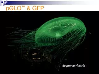
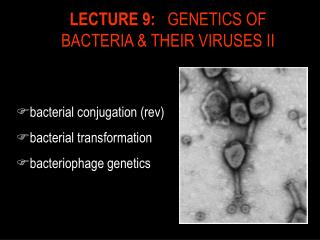
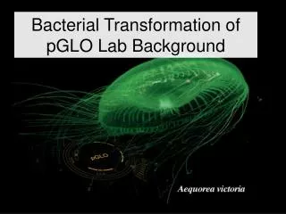
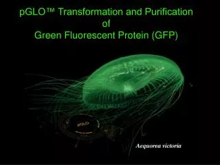
Discovery of GFP • Originally Isolated from the jellyfish Aequorea victoria • Naturally occurring in many bioluminescent jelly fish, reef corals and marine crustaceans • Recombinant GFP has 239 amino acids • Expressed as a 26,870 Dalton protein • Barrel structure with the fluorescent chromophore at center of the protein • “Nobel prize-winning” molecule!
What is a chromophore?A group of atoms and electrons forming part of an organic molecule that causes it to be colored The GFP chromophore is comprised of three adjacent amino acids.These amino acids are enzymatically converted to an active cyclic chromophore
GFP ChromophoreAbsorbs at 395 nmEmits at 509 nm • In vivo, GFP complexes with aequorin, a calcium-binding protein which transfers energy, to excite GFP • In vitro, UV light is used to excite the GFP chromophore, absorbing light at a wavelength of 395 nm, and emitting at a longer wavelength of 509 nm visible fluorescent green light
Recombinant GFP GFP has been mutated • Increased fluorescent photostability • Improved hydrophilicity • Increased solubility • Improved fluorescence • Various colors available • Improved use as a reporter protein
Using GFP as a biological tracer or reporter protein http://www.conncoll.edu/ccacad/zimmer/GFP-ww/prasher.html With permission from Marc Zimmer
Links to Real-world • GFP is a visual marker • Study of biological processes (example: synthesis of proteins) • Localization and regulation of gene expression • Cell movement • Cell fate during development • Formation of different organs • Screenable marker to identify transgenic organisms
Students will learn:Protein Electrophoresis of GFP • Prepare an SDS-PAGE sample and understand the components of Laemmli buffer • Understand protein structure and mechanisms for protein folding and unfolding and how different conformations can be identified using electrophoresis • Understand how proteins are separated during gel electrophoresis • Understand the use of electrophoresis in the process of transformation to protein expression • Understand chromophores and the basis of protein fluorescence • Construct a standard curve and determine the molecular weight (MW) of an unknown protein
Sample Preparation:SDS-Polyacrylamide Gel Electrophoresis (SDS-PAGE)“no heat” samplesLabeled tubes:1. White, no heat2. Green, no heat3. White, +heat4. Green, +heatSamples should be a “healthy scoop” of colonies • Label four screw-capped microtubes • Add 300 µl of Sample buffer to the two “no heat’ tubes • Using the inoculation loop, scrape a sample (20-100 colonies) from an LB/amp (white colonies) plate and transfer to the “White, no heat” tube and mix thoroughly • Using the inoculation loop, scrape a sample (20-100 colonies) from an LB/amp/ara (green colonies) plate and transfer to the “Green, no heat” tube and mix thoroughly
Sample Preparation SDS-PAGE“heat” samples • Transfer 150 µl of the “White, no heat” mixture to the “White,+heat” tube • Transfer 150 µl of the “Green, no heat” mixture to the “Green,+heat tube” • Heat the “+heat” tubes to 95°C for 5 min in a water bath. Cool to room temperature There is an important link between the STRUCTURE and FUNCTION of the protein Heating the samples denatures the proteins
CH3 CH2 CH2 CH2 CH2 CH2 CH2 CH2 CH2 CH2 CH2 CH2 O S O O - O SDS What is in the Laemmli sample buffer? • Tris buffer • provides appropriate pH • SDS (sodium dodecyl sulfate) • Solubilizes and denatures proteins • Adds a negative charge to the protein • DTT • (1,4-Dithiothreitol) reduces disulfide bonds, to help unfold proteins and protein complexes • Glycerol • Increases the density of the samples to help samples sink into wells of the gel • Bromophenol Blue • dye to visualize samples
Load and electrophorese samples30min at 200Vin 1XTGS Buffer UV illumination Coomassie Stain
s-s – + How Does SDS-PAGE Work? • Denatures proteins using detergent, DTT, and heat • Separates proteins based on size • Negatively charged proteins move to positive electrode • Smaller proteins move faster through the gel
Why Use Polyacrylamide Gels to Separate Proteins? • Polyacrylamide gel has a tight matrix • Ideal for protein separation • Smaller pore size than agarose • Proteins much smaller than DNA • Average amino acid = 110 daltons • Average nucleotide pair = 649 daltons • 1 kilobase of DNA = 650 kD • 1 kilobase of DNA encodes 333 amino acids = 36 kD • Size measured in kilodaltons (kD) • Dalton = mass of hydrogen molecule • = 1.66 x 10⁻²⁴ gram • Average amino acid = 110 daltons
GFP Visualization-During & Post Electrophoresis During Electrophoresis Post Electrophoresis • Fluorescent GFP can be visualized during electrophoresis • Coomassie stained gels allow for visualization of induced GFP proteins Fluorescent isoform Non- fluorescent isoform Prestained bands + UV activated GFP Coomassie stained bands
Determining the molecular weights of GFPs in different conformations
GFP Purification Procedures Overview Day 2 Day 1 Day 3
Why Use Chromatography? • To purify a single recombinant protein of interest from over 4,000 naturally occurring E. coli gene products.
Column Chromatography • Chromatography used for protein purification • Size exclusion • Ion exchange • Hydrophobic interaction
Hydrophobic Interaction Chromatography:(HIC)Steps 1–3 • Add bacterial lysate to column matrix in high salt buffer 2. Wash less hydrophobic proteins from column in low salt buffer • Elute GFP from column with no salt buffer
Hydrophobic bead H H O O - - - - H H + + O O O O S S N N H H H H O O + + O O - - - - O O S S O O - - O O + + Step 1:Hydrophobic Interaction Chromatography • Add bacterial lysate to column matrix in high salt buffer • Hydrophobic proteins interact with column • Salt ions interact with the less hydrophobic proteins and H2O
Hydrophobic bead H O - - + H O O S N H H O + + + + O - - - - - O S O - O + + + + Step 2:Hydrophobic Interaction Chromatography • Wash less hydrophobic from column with low salt buffer • Less hydrophobic E. coli proteins fall from column • GFP remains bound to the column
+ + + + + - - - - - + + + + + Step 3:Hydrophobic Interaction Chromatography Hydrophobic bead • Elute GFP from column by adding a no-salt buffer GFP • Released from column matrix • Flows through the column
Helpful Hints:Hydrophobic Interaction Chromatography • Add a small piece of paper to collection tube where column seats to insure column flow • Rest pipet tip on side of column to avoid column bed disturbance when adding solutions • Drain until the meniscus is just above the matrix for best separation
Column Chromatography vs. SDS-PAGEfor protein isolation and analysis Column Chromatography SDS PAGE
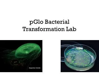
Transformationis only the beginning…… More techniques are necessary to fully understand the structure and nature of a protein. pGLO Transformation Protein Purification Results may lead to more experiments! Size and structure determination
Webinars • Enzyme Kinetics — A Biofuels Case Study • Real-Time PCR — What You Need To Know and Why You Should Teach It! • Proteins — Where DNA Takes on Form and Function • From plants to sequence: a six week college biology lab course • From singleplex to multiplex: making the most out of your realtime experiments explorer.bio-rad.comSupportWebinars