Cellular Responses to Heat and Oxidative Stress: Mechanisms and Transcriptional Regulation
370 likes | 482 Vues
This presentation covers key concepts in signal transduction and cellular responses to heat and oxidative stress. It explores the mechanisms by which regulatory proteins bind and function, particularly focusing on the heat shock response and its significance in transcription induction under stress conditions. Various proteins, including chaperones and proteases, play roles in managing misfolded polypeptides caused by temperature fluctuations. Additionally, we examine the role of sigma factors in transcription regulation and how cells sense temperature through chaperone concentrations.

Cellular Responses to Heat and Oxidative Stress: Mechanisms and Transcriptional Regulation
E N D
Presentation Transcript
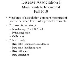
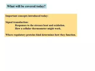
What will be covered today? Important concepts introduced today: Signal transduction: Responses to the stresses heat and oxidation. How a cellular thermometer might work. Where regulatory proteins bind determines how they function.
Cells induce transcription in response to various stresses in their environment. Table 3 shows some stresses and how many proteins are induced. (old data from 2-d gels) examples
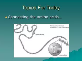
Heat shock response At 37 degrees they live happily in our gut. We get a fever and so do they. They produce proteins to counteract heat damage. See the proteins in the table, which deal with mis-folded polypeptides. They are mostly chaperones to help re-folding and proteases to degrade damaged proteins.
These will be relevant to regulation.
Heat shock response Array experiments show that these arise from increased transcription. How does heat signal increased transcription? And of course is there a thermometer? Analysis of promoters shows that they have in common a sequence that directs binding of the sigma32 form of holoenzyme.
Here are some of the transcription units that make heat shock proteins. They have no common repressor or activator. Instead their promoters have a unique sequence that binds polymerase with sigma32.
These sequences are found upstream of heat shock promoters Diversion: introduction into history of how elements are used by sigma factors.
TTGACA TATAAT A G 5 3 -35 region spacer -10 region +1 C T 5-8 bp Diversion - sigma70 and promoter elements Promoter alignment for 270 Consensus sequence has the most common nucleotide at each position. +10 • -20 • -40 • • • • 1st nuc. transcribed Is usually a purine 17+/- 1 TG AT-rich Analysis of promoter mutations 1st evidence for functional role of conserved sequences. The more similar the sequence is to the consensus, the higher is the RNA synthesis rate. This includes the 17 bps of the spacer length etc This allows appropriate (rather than maximal) RNA levels. -35 TTGACA TTTACA TTTACA “TATA” TATAAT TATGTT TATAAT consensus lac promoter lac UV5 (a stronger promoter)
This low resolution (cutaway) structure shows how sigma70 positions the DNA on the polymerase core. Other sigmas are expected to share this arrangement. Sigma region 4 binds -35 Sigma region 2 binds -10
Discuss now Discuss most other sigmas later
Back to heat shock response - is sigma the thermo-sensor? after hs (long-lived) amount of hot sigma32 before hs (t 1/2 of a minute) sigma32 protein pulse labeling experiment showed this result. Time of chase with cold amino acids Therefore, heat leads to accumulation of sigma32 Adam Blaszczak 1 , Costa Georgopoulos 2 & Krzysztof Liberek 3 Molecular Microbiology Volume 31ハIssue 1ハPage 157ハ - January 199
Isolation of complexes containing either of the 2 sigma factors implicates partner switching. (By gel filtration - see related exp in a few minutes) Partners soonafter heat shock: Partners before heat shock: For sigma32 - mostly polymerase (and it can transcribe) For sigma32 - mostly chaperones For sigma70 - mixture of chaperones and polymerase and aggregates of sigma itself For sigma70 - polymerase The next slide shows that association with polymerase is likely responsible for heat-induced protein stabilization.
Fts protease selectively degrades free sigma compared to core-bound Fig. 7 . RNA polymerase protects 32 from degradation by FtsH. [35S]- 32 (0.5 M) was incubated alone or with RNAP core (0.45 M) I n the presence of FtsH (0.28 M) in buffer A at 37°C. Molecular Microbiology Volume 31 Issue 1 Page 157 - January 1999
The KJE chaperones, which bind sigma before heat shock, do not protect sigma32 against degradation.
The next slide will put these data together into a model - Heat gives partner switching gives sigma32 activation
degraded + pol 70 Chaps P Chaps 32 Heat aggregation Misfolding etc 70 70 70 P* 70 Chaps pol 70 Chaps 32 Partner switch pol Chaps 32 Chaps 70 70 activation 70 70 P* 70
The GroE chaperone system is also involved Eric Guisbert1, Christophe Herman2,4,5, Chi Zen Lu2 and Carol A. Gross GENES & DEVELOPMENT 18:2812-2821, 2004 Just show 2 experiments that illustrate the binding of these to sigma32 and the inhibition of transcription.
Gel filtration shows that GroEL binds sigma32 (blot for sigma32) GroEL selectively inhibits sigma32 transcription free bound Note: KJ also inhibits sigma32 transcription Interpretation: Heat consumes another chaperone system, releasing sigma32 for transcription of heat shock genes. At the least this complements the KJE chaperone effects. Not known if one or the other dominates.
To sum up: is there a cellular thermometer? yes: temperature is measured by the concentration of free chaperone proteins. OK?
Here's another stress, oxidation damage, in this case from hydrogen peroxide. Bacteria can be oxidatively attacked by phagocytes in their preferred host. They can also live outside the host in toxic environments. Peroxide is one agent used to mimic such conditions. The next few slides just give a perspective on the oxyR regulon - General mechanisms of activation will come later.
Array experiments show that RNAs from the genes shown here are elevated by addition of hydrogen peroxide. Zheng et. al Journal of Bacteriology, August 2001, p. 4562-4570, Vol. 183
Many of these are proteins that are capable of reducing oxidized molecules of various sorts. The next slide will show that these constitute the oxyR regulon.
All promoters have oxyR binding sites. (and sigma70 consensus) Note: these views are downloaded from: http://BioCyc.org:1555//ECOLI/organism-summary
oxyR and peroxide are both needed to activate transcription in vivo.
And oxyR binds in vitro as part of activation. (DNase footprint)
Effects of peroxide Induction in vivo requires oxyR They depend on oxyR - A regulon
One notable exception to stimulation by oxyR - what’s different about the exception, which is the promoter that makes oxyR itself? up Transcription up up up up down
OxyR autorepression uses the usual active form of oxyR. Why does it repress, not activate? Old experiments on many systems show the answer to be: Location, location, location. Look at compilations to see zones of repression and activation.
Black bars show sites of repression; Others sites of activation. Note grouping of sites wrt +1 start of transcription
Location summary m-RNA -100 -80 -60 -40 -20 +1 +20 +40 Repression zone Activation zone And most activator proteins are really dual regulators as they sometimes repress by binding in the repression zone.
The dual regulators are the very same proteins in the same state when repressing or activating. In most cases only the location of the binding site has changed.
Why does location determine function? Constant pol footprint spacer -35 -10 m-RNA -100 -80 -60 -40 -20 +1 +20 +40 Repression zone Activation zone
Preview of activation mechanisms J. Mol. Biol (1998) 275:165 Each activator works from restricted positions within the activation zone (crp shown here).. If one artificially moves a natural site it activates best from places corresponding to natural positions (not shown). This is very different from repression and implies a need for stereospecific contact with pol. -42 -63 Natural locations of crp binding sites
Understanding repression zone in terms of closed and open complexes -40 -40 +20 +20 Repressors: Typically bind in repression zone (-40 to +20) and just block. Picture modified from Murakami see Science, (2002), 296, 1285-1290
Different zone for activators (more later) -40 -40 +20 +20 Activation zone - upstream from -40. They stabilize complexes when other elements are poor. Site phasing comes from need to contact a specific site on RNA polymerase. (eg work best on fixed side of DNA).
Homework assignment: This is figure 5 from Guisbert et al. GENES & DEVELOPMENT 18:2812-2821, 2004. Type one page describing why the 2 curves have the shapes shown.