Download
1 / 31
320 likes | 373 Vues
Learn about the functions of the kidney, nephron, renal blood supply, urinalysis tests, proteinuria, casts, hematuria, white blood cells, renal impairment tests, BUN, creatinine, NPN, urea, creatinine levels, and their clinical implications.

E N D
Kidney and Urinalysis Prepared by: Sr. Siti Norhaiza Hadzir
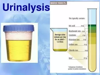
Functions of the kidney • Elimination of excess body water • Elimination of waste products of metabolism e.g urea & creatinine • Elimination of foreign substances e.g drugs • Retention of substances necessary for normal body function e.g protein, amino acids & glucose • Regulation of electrolytes balance & osmotic pressure of the body fluids.
The Nephron • The functional unit of the kidney. • Consists of renal corpuscle (glomerulus) & renal tubule. • Structure of glomerulus • Structure of tubule
Kidney blood supply • Renal artery from aorta → afferent arterioles → efferent arterioles → renal vein → heart
Glomerular Filtration Rate • Normally this amounts to about 130mL per minute (180 liters per 24 hours).
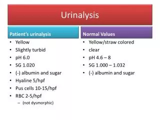
Renal Function Test • Falls into 2 major group: • Detect the presence of disease- not give indication as to the degree of functional impairment e.g proteinuria, cast, hematuria, WBC • Evaluate the degree of impairment e.g BUN, creatinine
Test of Urinary tract involvement • Healthy glomerular permeable membrane passes only substances with MW of less than 70 000. • Excess small proteins are reabsorbed completely by proximal tubule • Albumin is very close to cut off value (70000MW) can get access to the urine in glomerular disease. • Proteinuria are classified into 3: • Pre-renal- The glomerular membrane damage and tubular reabsorption inefficiency e.g Bence Jones protein in multiple myeloma. • Renal- renal parenchyma disease e.g amyloidosis. • Postrenal- Urinary tract problem e.g inflammation • Proteinuria
Figure 1: Normal urine is compared with proteinuria sample. Note increase in turbidity in proteinuria sample
Cast • Cast are precipitates of protein formed in the distal convoluted and collecting tubules of the kidney, where conditions of filtrate flow and pH are optimal for protein precipitation. • Normal condition-hyaline cast in small number • Large number indicates active renal disease.
Nature of cast • It is a muco-protein formed normally by the tubule; it is not formed in plasma. • It is long, rod like, flexible molecule. • As the glomerular filtrate travels down the nephron tubule, the concentrations of salts & H+↑. • At pH about 4.5, albumin and myoglobin change from negatively to positively charge molecules, the muco-protein is still negatively charge. • Opposite electric charge leads to precipitation and the formation of casts.
Hematuria & hemoglobinuria • Presence indicate bleeding within the urinary tract. • In acute glomerulonephritis there is hemorrhage from the glomeruli, Hb is convreted to hematin and methemoglobin. • These factors combine to give the “smoky” red brown urine characteristic of the disease.
Figure 2: The presence of blood in the urine
White Blood Cells • An increased number of white blood cells in a correctly collected specimen indicates inflammation in the urinary tract.
Test for Degree of Renal Impairment • Test based on water elimination and reabsorption • Blood Urea Nitrogen (BUN) • Creatinine • BUN: Creatinine
Test based on water elimination and reabsorption • Normally, conservation of water is reflected by concentrated urine with a high specific gravity • Excretion of an excess of water is illustrated by urine of low specific gravity
Impaired concentrating power • Tubular damage e.g chronic glomerulonephritis, polycystic disease • Severe potassium depletion • Hypercalcemia e.g due to vitamin D intoxication, hyperPTH • Inborn defects of tubular function • Diabetes insipidus
Non-protein nitrogen in blood • It is heterogenous collection of substances including urea, creatinine, uric acid, nucleotides, glutathione. • Estimation of NPN was replaced by determination of urea and creatinine, more specific indicators of renal condition, easily automated.
Blood Urea/BUN • Urea is the major excretion product of protein catabolism. • After elaboration, urea is passed to the blood and is excreted through the glomeruli and partly reabsorbed in the tubules.
Causes of ↑ BUN • Pre-renal: Circulation in the kidney is less efficient e.g CCF • Renal: Renal parenchyma damage, phylonephritis • Post-renal: Obstruction to the urinary tract • Presence of high level of urea is called uremia. • Very high level of urea leads to azotemia with kidney failure.
Creatinine • Nitrogenous substances found in muscle. • Since creatinine is derived entirely from endogenous metabolism (not form dietary protein) and is not reabsorbed by the renal tubules, its blood level; is a reliable index to renal function.
BUN/creatinine ratio • Normal ratio is 10:1. • Ratio more than 10:1 occur in: • Ration less than 10:1 occur in: • Excessive turnover of protein (hemorrhage, burns and infection) • Reduced glomerular perfusion • Repeated dialysis • Severe vomiting or diarrhea • Liver failure
Routine urinalysis • The procedure • Urine collection and storage • Macroscopic examination • - Color • - turbidity and clarity (smoky. milky, cloudy) • - smell • - SG and osmolality
Urine container Centrifuge tube Pipetting the supernatant
The procedure: cont; • Urine processing • Centrifuge • Separate debris and supernatant • Microscopic examination [cells (epithelium, RBC, • WBC, cast, mucus tread, ova and parasites, • crystals] • Biochemical analysis (pH, protein, glucose, ketones, bilirubin, blood, nitrite, urobinogen, ascorbic acid)