Structurally Mapping Membrane Proteins with Nuclear Magnetic Resonance
Learn how structural biologists at FSU are employing solid-state NMR to map the precise three-dimensional locations of proteins in cellular membranes. By aligning proteins in marching order, insights into complex protein structures are revealed.

Structurally Mapping Membrane Proteins with Nuclear Magnetic Resonance
E N D
Presentation Transcript
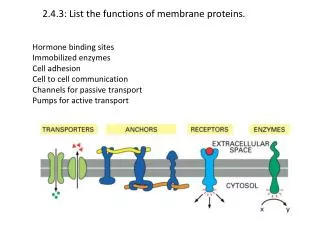
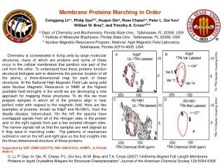
Conggang Lia,c, Philip Gaoa,c, Huajun Qina, Rose Chasea,c, Peter L. Gor’kovc William W. Breyc, and Timothy A. Crossa,b,c Membrane Proteins Marching in Order a Dept. of Chemistry and Biochemistry, Florida State Univ., Tallahassee, FL 32306, USA b Institute of Molecular Biophysics, Florida State Univ., Tallahassee, FL 32306, USA cNuclear Magnetic Resonance Program,National High Magnetic Field Laboratory, Tallahassee, Florida 32310-4005, USA Chemistry is orchestrated in living cells by large molecular structures, many of which are proteins and some of these occur in the cellular membranes that partition one part of the cell from the other. To understand how these proteins function structural biologists aim to determine the precise location of all the atoms, a three-dimensional map for each of these structures. At the National High Magnetic Field Lab using solid state Nuclear Magnetic Resonance or NMR at the highest available field strengths in the world we are developing a new approach for mapping these structures. To do this we must prepare samples in which all of the proteins align in near perfect order with respect to the magnetic field. Here are two examples of proteins, known as KdpF and Rv1861c, from the deadly disease, tuberculosis. On the left the spectra have overlapped signals from all of the nitrogen sites in the protein and on the right signals from just a few isolated nitrogen sites. The narrow signals tell us that the samples are well aligned as if they were in marching order. The patterns of resonances outlined in red on the left and right give us the first insights into the three dimensional structure of these proteins. Supported by NSF (DMB-0235774), NIH (GM-67476), NHMFL, & Florida State U. C. Li, P. Gao, H. Qin, R. Chase, P.L. Gor’kov, W.W. Brey and T.A. Cross (2007) “Uniformly Aligned Full-Length Membrane Proteins in liquid Crystalline Bilayers for Structural Characterization”Journal of the American Chemical Society 129:5304-5305