MICROSCOPES
MICROSCOPES. BY: Gabby Jones, Bridget Crawford, and Emily Baker. INDEX. Scanning Electron Microscope Atomic Force Microscope Electron Microscope. History Use Light Microscope Compound Microscope Scanning Tunneling Microscope X-Ray Microscope Binocular Microscope. History.


MICROSCOPES
E N D
Presentation Transcript
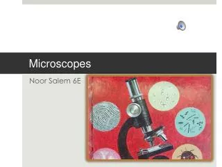
MICROSCOPES BY: Gabby Jones, Bridget Crawford, and Emily Baker
INDEX • Scanning Electron Microscope • Atomic Force Microscope • Electron Microscope • History • Use • Light Microscope • Compound Microscope • Scanning Tunneling Microscope • X-Ray Microscope • Binocular Microscope
History The first record of the use of lenses to manipulate images was in Greek and Roman writings of around 1000 A.D. As for the origins of someone using lenses to magnify a minute object, it is unclear. Most scientific instruments have a clear place in the historical records when they were formed and who created them, not the microscope though. The definition of the microscope makes it difficult to determine when it was first created. Since there were lenses dating back to ancient societies, how do we say when those lenses were used to look at minute objects? It is practically impossible to say when a single lens was used in that fashion. Credit for the first compound microscope (multiple lenses) is generally given to Zacharias Jansen and John Lipperhey of the Netherlands, in 1590.
USE • A microscope is an instrument that produces an enlarged image of an object.
Light Microscope • A microscope ( Greek: Micran= small and scopos = aim) is an instrument for veiwing objects that are to small to be seen by the naked or unaided eye. The science of investing small objects using such an instrument is callled microscopy, and the term microscopic means minute or very small, not easily visible with the unaided eye. In other words, requiring a microscope to examine.
Compound Microscopes A microscope which consists of two microscopes in series, the first serving as the ocular lens (close to the eye) and the second serving as the objective lens (close to the object to be viewed). Credit for creating the compound microscope goes usually to the Dutch spectacle makers Hans and Zacharias Janssen who in 1590 invented an instrument that could be used as either a microscope or telescope. The compound microscope has evolved into the dominant type of optical microscope today.
Scanning Tunniling Microscopes • The scanning tunneling microscope (not to be confused with scanning electron microscopes electron microscopes), or STM, was invented in 1981 by Gerd Binning and Heinrich Rohrer of IBM's Zurich Lab in Zurich, Switzerland. Although initially greeted with considerable scepticism by materials scientists in the early 1980s, the invention garnered the two a Nobel Prize in Physics in 1986. The STM allows scientists to visualize regions of high electron density and hence infer the position of individual atoms, where previously arduous study of diffraction patterns from prior methods lead to much debate as to the real, spatial lattice structure of the item in question. The STM has higher resolution than its slightly later invented but nevertheless related cousin, the atomic force microscope (AFM). Both the STM and the AFM fall under the class of scanning probe microscopy instruments. • It is used to obtain images of conductive surfaces at an atomic scale 2 × 10⁻¹⁰ m or 0.2 nanometre. It can also be used to alter the observed material by manipulating individual atoms, triggering chemical reactions, and creating ions by removing individual electrons from atoms and then reverting them to atoms by replacing the electrons.
X-Ray Microscopes An X-ray microscope uses electromagnetic radiation in the soft X-ray band to produce images of very small objects. Unlike visible lightmicroscopes, X-rays do not reflect or refract easily, and they are invisible to the human eye. Therefore the basic process of an X-ray microscope is to expose film or use a charge-coupled device (CCD) detector to detect X-rays that pass through the specimen, rather than light which bounces off the specimen. Early X-ray microscopes by Kirkpatrick and Baez used grazing-incidencereflective optics to focus the X-rays, which grazed X-rays off parabolic curved mirrors at a very high angle of incidence. An alternative method of focusing X-rays is to use a tiny fresnelzone plate of concentric gold or nickel rings on a silicon dioxide substrate. Sir Lawrence Bragg produced some of the first usable X-ray images with his apparatus in the late 1940's. In the 1950's Newberry produced a shadow X-ray microscope which placed the specimen between the source and a target plate, this became the basis for the first commercial X-ray microscopes from the GeneralElectric Company.
Binocular Microscopes • A compound microscope with two eyepieces viewing down a single optical channel and objective lens. This is different than a stereo microscope, which has a separate optical channel for each eye.
Scanning Electron Microscopes
Atomic Force Microscope • The Atomic Force Microscope (AFM) is a very powerful microscope invented by Binnig, Quate, and Gerber in 1986. Besides imaging it is also one of the foremost tools for the manipulation of matter at the nanoscale.
Electron Microscope • The electron microscope is a microscope that can magnify very small details with high resolving power due to the use of electrons rather than light to scatter off material, magnifying at levels up to 500,000 times