
Pathogenic Spirochetes: Classification and Implications
E N D
Presentation Transcript
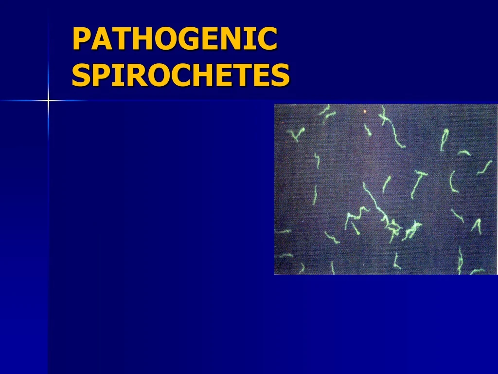
Introduction: • Spirochetes: slender, spiral, motile, flexible gram negative bacilli • speira= coil, chaite= hair • Two families: • Spirochetaceae • Leptospiraceae
Taxonomy • Order: Spirochaetales • Family: Spirochaetaceae • Genus: Treponema • Borrelia • Family: Leptospiraceae • Genus: Leptospira
Spirochetes • Treponema • Borrelia • Leptospira
T pallidum has an outer sheath (glycosaminoglycan coating) • Inside the sheath is: outer membrane (peptidoglycan) (maintains the structural integrity) • Endoflagella (axial filaments) in the periplasmic space encased by the outer membrane • Endoflagella begin at each end of the organism, wind around it, extending to and overlapping at the midpoint.
Inside the endoflagella is the inner membrane (cytoplasmic membrane): provides osmotic stability and covers the protoplasmic cylinder. • A series of cytoplasmic tubules (body fibrils) are inside the cell near the inner membrane. They reproduce by transverse fission.
Spirochetes ultrastructure Borrelia Leptospira
Spirochetes morphology Treponema Leptospira Borrelia
Treponema Pathogenic Non-pathogenic Oral commensals Pathogenic Non-venereal disease by direct contact T. Denticula T.macrodentium T.microdentium T.pallidum Syphilus T.Pertenue T. carateum T.pallidum A sexually transmitted disease Yaws Pinta Bejal
Non-pathogenic T.denticola T.macrodenticum T.orale Conditionally pathogenic T.vincentii Pathogenic T.pallidum pallidum (syphilis) T.pallidum endemicum (bejel) T.pallidum pertenue (yaws) T.carateum (pinta) Classification of Human Treponemes
S Y P H I L I S Causative Organism – Treponema pallidum
Characteristics of T.pallidum • T.pallidum: motile, slender • Culture: Pathogenic T.pallidum has never been cultured continuously on artificial media • Non-pathogenic treponemes : Reiter strain can be cultured anaerobically in vitro, on a defined medium of 11 amino acids, vitamins, salts, minerals, and serum albumin. • T. pallidum microaerophilic organism; survives best in 1–4% oxygen • Propagated by inoculation in rabbits testes & anterior chamber of eye.
Can be seen by • Dark field microscopy • Phase contract technique • Can be stained by • Silver impregnation • Fluorescent antibody technique • Sensitive to penicillin
Reactions to Physical and Chemical Agents: • Drying & temperature to 42°C kills the spirochete rapidly • Penicillin is the drug of choice
Pathogenesis of T. pallidum • Tissue destruction and lesions are primarily a consequence of patient’s immune response • Syphilis is a disease of blood vessels and of the peri-vascular areas • In spite of a vigorous host immune response the organisms are capable of persisting for decades • Infection is neither fully controlled nor eradicated • In early stages, there is an inhibition of CMI • Inhibition of CMI abates in late stages of disease, hence late lesions tend to be localized
Virulence Factors of T. pallidum • Outer membrane proteins: Adherence • Hyaluronidase: for peri-vascular infiltration • Antiphagocytic coating of fibronectin • Tissue destruction and lesions are primarily result of host’s immune response (immunopathology) • Cardiolipin is an important component of the treponemal antigens.
Mode of Transmission • Direct sexual contact (90 – 96%) • Blood transfusion • Congenital syphilis: Via placenta from infected pregnant mother fetus causes • Accidental contact: Medical personnel. Source of T.pallidum: Primary and secondary syphilis lesions Incubation Period: 10 – 90 days (average: 21 days)
Acquired Syphilis • Natural infection with T pallidum is limited to the human host. • Human infection usually sexually transmitted • Infectious lesion on skin or mucous membranes of genitalia. • In 10–20% of cases, however, the primary lesion is intra-rectal, perianal, or oral. • T pallidumpenetrates intact mucous membranes, or enters through a break in the epidermis.
Stages of Disease • Primary • Secondary • Latent • Tertiary • Congenital Syphilis
Primary syphilis- Spirochetes multiply locally at the site of entry, spread to local lymph nodes, reach the bloodstream. • In 2–10 weeks after infection, a papule develops at the site of infection and breaks down to form an ulcer with a clean, hard base ("hard chancre“) which is painless • Primary lesion: predominance of lymphocytes and plasma cells. "primary lesion“ heals spontaneously, but 2–10 weeks later the "secondary" lesions appear.
Fluid from chancre is highly infectious • Dark field microscopy shows spirochetes. • There is regional lymphadenopathy. • Primary chancre heals spontaneously in 3-8 weeks. • Primary syphilis is highly infectious. • Serological tests for Syphilis are positive in 80% cases.
Secondary Syphilis • Occurs 6-8 weeks after initial chancre, patient highly infectious. • Characterized by localized or diffuse mucocutaneous lesions, with generalized lymphadenopathy. • Primary chancre may still be present. • Secondary lesions subside in about 2-6 weeks. • Serology tests nearly 100% positive.
Latent Stage: • After the secondary syphilis symptoms subsides, the disease enters a latent stage. • Stage of infection in which organisms persist in the body without symptoms or signs(asymptomatic). • 1/3rd of untreated latent stage individuals develop tertiary syphilis • After about 2 years, the syphilis is NOT normally infectious, except from mother to the foetus.
Tertiary Syphilis • Three manifestations: • Gummatous syphilis • Cardiovascular syphilis (aortic aneurysms) • Neuro syphilis: Generalized paresis of the insane Tabesdorsalis
Tertiary Syphilis - Gummatous • Gummas: localized areas of granulomatous inflammation found on bones, skin and s/c tissue • Cutaneous gummas: single or multiple, generally asymmetric and grouped together. • Visceral lesions: cause local destruction of affected organ. • Contain lymphocytes, plasma cells and perivascular inflammation.
Neuro syphilis: • Frontal lobe Cerebral atrophy seen in general paresis • Generalized paresis of the insane:personality changes, emotional affect, hyperactive reflexes • Tabesdorsalis: degeneration of lower spinal cord, general paresis, chronic progressive dementia often results in a shuffling gait • Neurosyphilis can only be diagnosed serologically by VDRL.
Congenital Syphilis • Transmitted from mother to fetus. • Fetus affected during second or third trimester • 40-70% infected, 12% will subsequently die prematurely • Bone deformities • Blindness (Interstitial keratitis, blindness), Deafness • Deformed face: saddle nose • Dental deformities (Hutchinson’s incisors, Mulberry molars) • Skin rashes (rash on soles & palms) • Hepato-spleenomegaly, Lymphadenopathy • Mental retardation, Stillbirths
Mulberry Molars Hutchinson’s incisors
Diagnosis of Syphilis • Evaluation based on three factors: • Clinical findings. • Demonstration of spirochetes in clinical specimen. • Present of Abs in blood or CSF • More than one test should be performed.
Diagnostic Tests for Syphilis (Original Wasserman Test) NOTE: Treponemal antigen tests indicate experience with a treponemal infection, but cross-react with antigens other than T. pallidum ssp. pallidum. Since pinta and yaws are rare in USA, positive treponemal antigen tests are usually indicative of syphilitic infection.
Dark ground microscopy: To demonstrate, Spirochaetes T. pallidum in fluid or exudate from lesions of primary and secondary syphilis. a) Primary Syphilis: exudate from chancre b) Secondary syphilis: mucous patch exudate • Direct immuno-fluorescent microscopy can be used.
Non-treponemal Reagin Tests • Serological test to detect reaginAb • Screening test only • Reagin: Ab formed against cardiolipin Ag • Found in sera of patients with syphilis as well as other diseases. • Non treponemal tests: positive 1-4 wks after appearance of primary chancre.
Nontreponemal Reagin Tests • VDRL • Wasserman raection • RPR • RST-reagin screen test
Venereal Disease Research Laboratory - VDRL • Slide Flocculation test (serum & CSF) • VDRL Ag: consists cardiolipin, cholesterol, lecithin. • Must be made up fresh daily. • Serum inactivation: heated to 560 C for 30 min to remove anti-complementary activity which may cause false positive • Calibrated syringe utilized to dispense antigen
Rapid Plasma Reagin Test - RPR • Screening test • CANNOT be done on CSF • Antigen • VDRL cardiolipin antigen is modified with choline chloride to make it more stable • Charcoal particles added: Macroscopic reading • Serum or plasma may be used for testing, serum need not be activated