Download
1 / 15
150 likes | 356 Vues
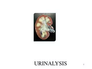
Biology 161 - Urinalysis. Scott.lehbauer@lethbridgecollege.ab.ca. Kidney Structures. Renal Artery. Renal Vein. Renal Capsule. Ureter. Kidney Structures. Glomerulus. Renal Calyx. Renal Pyramid. Renal Sinus. Kidney Structures. Renal Cortex. Renal Medulla. Kidney Structures.

E N D
Biology 161 - Urinalysis Scott.lehbauer@lethbridgecollege.ab.ca
Kidney Structures Renal Artery Renal Vein Renal Capsule Ureter
Kidney Structures Glomerulus Renal Calyx Renal Pyramid Renal Sinus
Kidney Structures Renal Cortex Renal Medulla
Kidney Structures Renal Pyramid Renal Calyx Renal Capsule Renal Cortex
Kidney Structures Renal Capsule (Covering) Renal Hilum
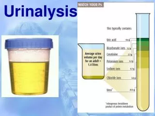
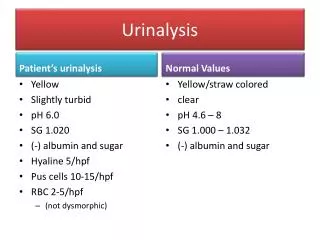
Urinalysis 1.) Test for pH (Bayer Sticks) • Normal pH of urine is 4.5 – 8.0 2.) Color and Turbidity • Normal urine is pale yellow and clear
Urinalysis 3.) Specific Gravity of Urine – specific gravity compares the weight of the urine to the weight of water. • Water is assigned a specific gravity of 1.00 • Urine has a normal specific gravity of 1.003 to 1.035 • High specific gravity indicates dehydration • Low specific gravity may indicate diabetes insipidus.
Urinalysis Specific Gravity Cont. How to Read the urinometer • Fill the cylinder ¾ full of urine. • Place the hydrometer into the urine. • Read the scale at the top of the urine. Midget Urinometer
Urinalysis 4.) Reading the multistix. • Completely insert the multistix into the urine. • Read multistix from top to bottom on the chart. Note – Do not let the multistix touch the chart itself while reading it
Urinalysis Possible Multistix Positives • Blood – may occur with burns, crushing injuries, hemolytic anemia. • Ketones – may occur with diabetic acidosis. • Glucose – may occur with diabetes mellitus or with extra intake of sugar. • Protein – may occur with chronic renal failure.
Urinalysis • The pH – should be in the 4.5 to 8.0 range • Bilirubin – increase with bile duct blockages • Urobilinogen – with total obstruction of bile flow, no bilirubin reaches the intestines to be worked on by bacteria and changed to urobilinogen in the urine. Urine tends to be pale since urobilinogen colors the urine.
Urinalysis Benedict’s – tests for the presence of a monosaccharide (reducing sugar) or in our bodies case glucose. The solution will turn from blue to brick red in the presence of glucose.
Urinalysis Biuret – Tests for the presence of protein in solution. In the presence of protein the solution will turn from blue to violet in color.
Urinalysis Sulfur – will test for the presence of bile in the urine. If the pinch of sulfur floats in the urine then there is no bile present. If the sulfur sinks and becomes frothy then there is bile present in the urine.