BODY PLANES, DIRECTIONS, AND CAVITIES
250 likes | 1.18k Vues
BODY PLANES, DIRECTIONS, AND CAVITIES

BODY PLANES, DIRECTIONS, AND CAVITIES
E N D
Presentation Transcript
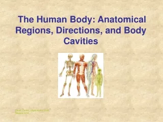
BODY PLANES, DIRECTIONS, AND CAVITIES Understanding anatomical directional terms and body planes will make it easier to study anatomy. It will help you to be able to visualize positional and spacial locations of structures and navigate directionally from one area to another. Another strategy that can be employed to help you visualize anatomical structures and their positions is to use study aids such as anatomy coloring books and flash cards. It may seem a bit juvenile, but coloring books and review cards actually help you to visually comprehend the information.
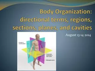
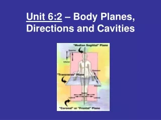
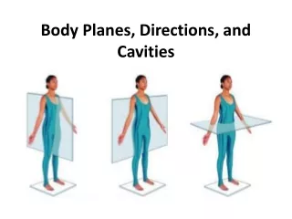
Anatomical Body Planes: Imagine a person standing in an upright position. Now imagine dissecting this person with imaginary vertical and horizontal planes. This is the best way to describe anatomical planes. Anatomical planes can be used to describe any body part or an entire body.
A plane is a two-dimensional surface — its dimensions are length and width. The body reference planes are used to locate or describe the location of structures in the body. These terms are often used to describe medical imaging such as CAT scans, PET scans and MRIs where the scans take pictures of the body in flat slices. Brain scans are often of sagittal plane slices (from ear to ear). Abdominal CAT scans are often transverse plane slices (like a stack of coins).
Transverse Plane: Imagine a horizontal plane that runs through the midsection of your body. This plane divides the body into upper (superior) and lower (inferior) regions. Frontal Plane or Coronal Plane: Imagine a vertical plane that runs through the center of your body from side to side. This plane divides the body into front (anterior) and back (posterior) regions. Sagittal Plane: Imagine a vertical plane that runs through your body from front to back or back to front. This plane divides the body into right and left regions. Midsagittal Plane: Sagittal plane that divides the body into equal right and left regions.
Directional Terms: In general, directional terms are grouped in pairs of opposites based on the standard anatomical position. Superior and Inferior. Superior means above, inferior means below. The elbow is superior (above) to the hand. The foot is inferior (below) to the knee. Anterior and Posterior. Anterior means toward the front (chest side) of the body, posterior means toward the back. Medial and Lateral. Medial means toward the midline of the body, lateral means away from the midline. Ipsilateralmeans on the same side—the left arm is ipsilateral (on the same side) to the left leg. Proximal and Distal. Proximal means closest to the point of origin or trunk of the body, distal means farthest away. Proximal and distal are often used when describing arms and legs. If you were describing the shin bone, the proximal end would be the end close to the knee and the distal end would be the end close to the foot. In the fingers of the hand, a proximal joint is closest to the wrist and a distal joint is farthest from the wrist. Superficial and Deep. Superficial means toward the body surface, deep means farthest from the body surface. Cranial and Caudal. Cranial means Towards the head, Caudal means toward the back or tail
Quadrants divide our bodies into regions for diagnostic and descriptive purposes. The quadrants are defined by drawing an imaginary line vertically (top to bottom) and horizontally (sideways) though the umbilicus (belly button). Right Upper Quadrant (RUQ) – right lobe of liver, gallbladder, part of the transverse colon, part of pylorus, hepatic flexure, right kidney, and duodenum. Right Lower Quadrant (RLQ) – cecum, ascending colon, small intestine, appendix, bladder if distended, right ureter, right spermatic duct (men), right ovary and right tube and uterus if enlarged (women). Left Upper Quadrant (LUQ)– Left lobe of liver, stomach, small intestine, transverse colon, splenic flexure, pancreas, left kidney and spleen. Left Lower Quadrant (LLQ) – small intestine, left ureter, sigmoid flexure, descending colon, bladder if distended, left spermatic duct (men) left ovary and left tube and uterus if enlarged (women).
Body regions describe areas of the body that have a special function or are supplied by specific blood vessels or nerves. The most widely used terms are those that describe the 9 abdominal regions shown in the image to the right. The regions are named below and the corresponding regions are labeled 1-9. Abdominal Regions right (1) and left (3) hypochondriac regions– on either side of the epigastric region. Contains the diaphragm, some of the kidneys, right side of the liver, the spleen and part of the pancreas. epigastric region (2)– superior (above) the umbilical region and contains most of the pancreas, part of the stomach, liver, inferior vena cava, abdominal aorta and duodenum right (4) and left (6) lumbar (lateral) regions– on either side of the umbilical region. They contain portions of the large and small intestines and kidneys. umbilical region (5) – area around the umbilicus (belly button). Includes sections of the large and small intestines, inferior vena cava and abdominal aorta right (7) and left (9) iliac (inguinal) regions– are on either side of the hypogastric region and include portions of the large and small intestines. hypogastric (pubic) (8) region– inferior (below) the umbilical region. Contains parts of the sigmoid colon, the urinary bladder and ureters, the uterus and ovaries (women), and portions of the small intestines.
Body cavities are Spaces within the body that contain the internal organs. Dorsal Body Cavity • Cranial Cavity • Contains the brain • Spinal (Vertebral) Cavity • Bony cavity formed by the vertebrae of the spine that contains and protects the spinal cord.
Ventral Body Cavity • Thoracic Cavity • Pleural cavities (2) • Mediastinum • Pericardial cavity • Abdominopelvic Cavity • Abdominal cavity • Pelvic cavity
Review Question 3 Nose - Calf of Leg - Ears - Umbilicus - Fingernails – Possible answers: Nasal, navel, Otic, Sural, Digital, gluteal, buccal
Answers: • A. Foot • B. Cheekbone • C. Chin • D. Toenails • E. Skin
Answer: • Frontal or Coronal
Answers to At the Clinic 1 • Antecubital – anterior surface of elbow • Deltoid Region – Curve of shoulder formed by large deltoid muscle, Took off his shirt • Sural Region - Calf
Answers to clinic 2 • Axcillary – Armpit • Cervical – Neck • Scapular – Shoulder Blade • Brachial – Arm bend
Test next class on ch 7:2 worksheets, Power point, and body landmarks • STUDY!!