Download

1 / 44
450 likes | 603 Vues
An Introduction to. Body Planes, Directions, Cavities, and Regional Terminology. Introduction. LMAO!. Anatomic reference systems describe the location and functions of body parts. The basic reference systems are: body planes body directions body regions body cavities. Objectives.

E N D
An Introduction to Body Planes, Directions, Cavities, and Regional Terminology
Introduction LMAO! • Anatomic reference systemsdescribe the location and functions of body parts. The basic reference systems are: • body planes • body directions • body regions • body cavities
Objectives • understand the how 3 body planes divide the human body • Be able to use directional terminology in describing different areas of the body • Identify and recognize body regions • Gain a working understanding of body cavities and the organs they house
Anatomical Position • Person stands erect with feet together and eyes forward • Palms face anteriorly with thumbs pointed away from the body • Right and left always refers to the sides belonging to the person or specimen being viewed – never to the viewer • Note: four legged animals have a different anatomical position than humans • Their ventral is on the inferior side and dorsal in on the superior side • In humans ventral and anterior is the same and so is dorsal and posterior
Directional Terminology 1. Anterior = body parts on the front of the body 2. Posterior = body parts on the back of the body The frontal plane divides the body into “anterior” and “posterior” regions.
Directional Terminology • Cranial or Superior = body parts near the head • Caudal or Inferior = body parts located near the sacrum, or tail bone. Cranial Caudal
Directional Terminology • Medial = body parts located near the middle or midline of the body 2. Lateral = body parts located away from the midline or middle of the body
Lateral and Medial • Lateral referrs to
Directional Terminology • Proximal = body parts close to the point of reference • Distal = body parts away from the point of reference
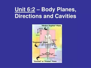
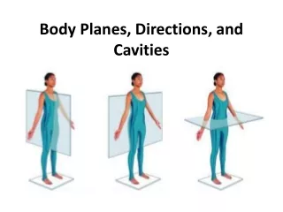
What is a “Plane?” • A “body plane”is an imaginary line drawn through the body which separates it into sections.
Body Planes: The Sagittal Plane -The Sagittal Plane Divides the body into right and left sides
Body Planes: The Frontal Plane The “Frontal Plane” divides the body into front and back section. The frontal plane is sometimes called the “Coronal Plane.”
Body Planes: The Transverse Plane • The “transverse Plane” divides the body into sections above and below the midline.
The Orange activity • Use a marker to label the top and bottom of your orange. • Draw a line around the orange which represents the transverse plane. Label. • Draw a line around the orange which represents the frontal or coronal plane. Label. • Draw a line around the orange which represents the Sagittal Plane. Label.
The Orange activity • Cut the orange in half along the transverse plane. When Finished, use a toothpick to put the orange back together. • Cut the orange in half along coronal plane. When Finished, use toothpicks to put the orange back together. • Cut the orange in half along the Sagittal Plane. When Finished, use toothpicks to put the orange back together.
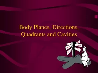
Abdominal Regions: Quadrants 1. Right Upper Quadrant (RUQ) 2. Left Upper Quadrant (LUQ) 3. Right Lower Quadrant (RLQ) 4. Left Lower Quadrant (LLQ)
Abdominal Regions: • Epigastric • Umbilical • Pelvic • Hypochondriac • Lateral • Inguinal
Body Cavities: The Dorsal Cavity -A long continuous cavity that is located on the back (or posterior) of the body, divided into two sections Cranial Cavity = contains the brain Spinal Cavity = contains the spinal cord
Divisions of the Spinal Cord • Cervical vertebrae: C • Thoracic vertebrae: T • Lumbar vertebrae: L • Sacrum: S
Body Cavities: The Anterior Larger and separated into 2 distinct cavities by a dome-shaped muscle called the diaphragm, which is important for breathing. • Thoracic Cavity = located in the chest, contains the heart, lungs, and the large blood vessels
Pericardial, Pleural, Peritoneal Figure 1.7
Body Cavities: The Anterior 2. Abdominal Cavity = divided into quadrants… Upper part contains the stomach, small intestines, most of the large intestines, liver, gallbladder, pancreas and spleen 3. Pelvic Cavity = lower abdominal cavity containing urinary bladder, the reproductive organs, and last part of the large intestines
Body Cavities Cranial Thoracic Spinal Diaphragm Adbdominal Pelvic
Clinical Anatomy: An Introduction to Medical Imaging Techniqes Traditional more non-invasive method of diagnosis • X-rays (electromagnetic waves) directed at the body • Some x-rays are absorbed: amount of absorption depends on the density of matter encountered • Radiograph image: negative • Darker exposed areas represent soft organs (easily penetrated) • Light, unexposed areas correspond to denser structures such as bones • Contrast medium: solution with heavy elements (i.e. barium) • Used to view soft tissue organs • Advanced X-Ray techniques use computer-assisted imaging technologies
Radiography • X ray: electromagnetic waves of very short length • Best for visualizing bones and abnormal dense structures Clavicles (collarbones) Ribs Air in lungs (black) Heart Diaphragm (a) Radiograph of the chest (b) Mammogram (cancerous tumor at arrow) Figure 1.10
Advanced Medical Imaging: Computed (Axial) Tomography (CT or CAT) Right Left • Takes successive X rays around a person’s circumference • Translates recorded information into a detailed picture of the section Liver Stomach Colon View Inferior vena cava Aorta Spleen Left kidney Thoracic vertebra
Contrast X-rays • Contrast media make hollow or fluid-filled structures visible • Media can be introduced by injection, orally, or rectally • Depends on the structure imaged Barium contrast x-ray showing a cancer of the ascending colon (arrow)
Digital Subtraction Angiography (DSA) • A contrast medium given: images taken ‘before’ and ‘after’ • Computer processes the x-ray images and subtracts the differences • Eliminates all traces of body structures that obscure the vessel • Identify blockages of arteries that supply the heart or brain Figure 1.12
PET (Positron Emission Tomography): accesses functional flow of blood to the heart & brain • Produces images by detecting radioactive isotopes injected into the body • Decaying isotopes emits gamma rays • Detected by sensors, translated into impulses and sent to a computer • Active areas receiving more blood light up Figure 1.13
Sonography (Ultrasound Imaging) • Pulses of high frequency (ultrasonic) sound waves reflect (echo) off tissue • Computer analyzes the echoes to construct sectional images • Inexpensive/safer technique but not used for viewing air-filled structures or structures surrounded by bone Figure 1.14
Magnetic Resonance Imaging (MRI) • High-energy magnetic field causes protons (H+) in tissues and fluids to align in relation to the field • Pulse of radio waves emitted to misalign H+ • As they realign with the magnet a radio wave is again emitted • Sensors ‘read’ these ion patterns, computerized signals produce detailed images of soft tissues
Endoscopy • Endoscope: lighted instrument with lenses • Used for visual examination of the inside of body organs or cavities • Colonoscopy: interior of the colon • Arthroscopy: interior of a joint • Laparoscopy: interior of abdominopelvic organs Interior view of the colon as shown by colonoscopy