Practical No.14 Genus Bacillus
190 likes | 208 Vues
This practical article discusses the genus Bacillus, specifically Bacillus anthracis, the causative agent of anthrax, and Bacillus cereus, which causes foodborne illness. It covers the symptoms, diagnostic tests, and treatment options for these diseases.

Practical No.14 Genus Bacillus
E N D
Presentation Transcript
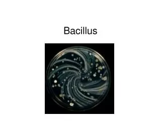
Practical No.14 Genus Bacillus
Genus Bacillus Saprophytic spp. Pathogenic species Bacillus anthracis Bacillus subtilis Bacillus cereus Animal reservoir Dust, soil, water… Anthrax Conjunctivitis Food poisoning
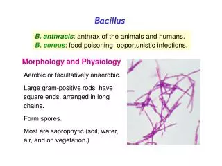
G+ve aerobic spore-forming bacilli: Two Bacillus species are considered medically significant: B. anthracis, which causes anthrax, and B. cereus, which causes a foodborne illness. A third species isB. subtilis, an important model organism. It is also a notable food spoiler; it is also one of the contaminants of culture media in bacteriology laboratories.
Bacillus anthracis: Bacillus anthracis is a Gram-positive spore-forming, rod-shaped bacterium. Bacillus anthracis spores in particular are highly resilient, surviving extremes of temperature, low-nutrient environments, and harsh chemical treatment over decades or centuries. It is the causative agent of anthrax. Three types of anthrax: 1. Cutaneous anthrax (malignant pustule). 2. Pulmonary anthrax (wool sorter's disease) and 3. Gastrointestinal anthrax.
Bacillus anthracis diseases: Cutaneous anthrax (95% of cases; malignant pustule (contact with infected animals or their products, spore, biting fly). Inhalation anthrax (4% of cases; Wool sorter's disease) . (Inhalation of spore from the dust of wool, hair, or hides ) Gastrointestinal anthrax (rare) .
Laboratory diagnostic tests: Specimen: pus, sputum, stool. Smear:stained with gram's stain.Appearsas large G +ve bacilli arranged singly, pairs or in chains with squared ends. In laboratory media, the bacilli form long chains. Spores develop at the end of the log phase of multiplication. These are central / subterminal, ellipsoidal and non-bulging and resist Gram stain, appearing as unstained areas within the cell. With malachite green/safranine (or malachite green/ basic fuchsin) staining, the spores are stained green and the vegetative forms are pink. In the Ziehl-Neelsen staining, spores are pink and the vegetative forms are blue. Capsules are lost in normal culture, the capsule is only seen if the specimen is taken from animal's exudates or from blood, by using a Mcfadyean reaction, capsule appears reddish purple and the bacillus appear blue. Mcfadyean reaction involves staining with polychrome methylene blue.
Specimens: Cutaneous anthrax Fluid (aspirate) or pus from lesion. Inhalation anthrax Sputum. Staining: -Gram stain: G + ve bacilli, cutted ends, arrange in chains, colorless spore. -Capsular staining: bacillus surrounded by halo (capsule). Capsular staining for Bacillus anthracis
Microscopical Characteristics G +ve, bacilli Cutted ends Arranged either single, pairs but mainly in chains bulging spore
Culture:on blood agar and under aerobic conditions colonies appear white, granular, circular disks which have margins resembling a lock of hairs (the medusa head). • Biochemical reactions:gelatin liquification ; Bacillus anthracisgives a positive result.
Macroscopical Characteristics:- Aerobic. Haemolysis Bacillus anthracis gray medusa head (under low power microscope), ground glass (frosted glass) appearance Saprophytic spp. circular with irregular margins Non – motile Bacillus anthracis Motile Saprophytic spp.
Bacillus subtilis on blood agar Showing hemolysis, irregular margin due to motility
Gelatin liquefaction (A) Negative. (B & C) positive
Differential characteristics of B. anthracisand non-pathogenic B. species
Serological tests: Ascoli precipitin test; is used for recognition of anthrax infection. About 2 grams of tissue is boiled for 5 minutes with 5 ml normal saline to which acetic acid has been added. The fluid is filtered, 0.5 ml of anthrax antiserum is placed in a narrow tube and clear filtrate is carefully layered on the top of the serum. The development of a white ring of precipitate at the junction of the two fluids within 10 minutes at room temperature, denote positive results.
Serological Tests ELISA, IFA monoclonal antibodies against bacteria or exotoxin MAINLY USED NOW PCR Ascoli’s thermo-precipitatin : ring precipitation test to demonstrate anthrax Ag in tissue extract. • Antibiotic sensitivity: • Bacillus anthracisis SENSITIVE to penicillin. • saprophytic spp. are RESISTANTto it.