Ch 7 – The Microscope
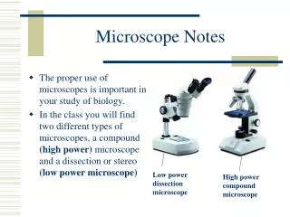
Ch 7 – The Microscope. Compound microscope. Magnification, field of view, working distance, and depth of focus. Comparison microscope. Advantages of stereoscopic microscope. Plane-polarized light and polarizing microscope. Advantages of linking a microscope to a spectrophotometer.

Ch 7 – The Microscope
E N D
Presentation Transcript
Ch 7 – The Microscope • Compound microscope. • Magnification, field of view, working distance, and depth of focus. • Comparison microscope. • Advantages of stereoscopic microscope. • Plane-polarized light and polarizing microscope. • Advantages of linking a microscope to a spectrophotometer.
Utilizing a microspectrophotometer for examining trace physical evidence. • Mechanism of image formation for light microscope Vs scanning electron microscope (SEM). • Advantages and applications of SEM in forensic science.
Virtual image: an image cannot be seen directly. It can only be seen by a viewer looking through a lens. • Real image: an image formed by the actual convergence of light rays upon a screen • Objective lens: the lower lens of a microscope that is positioned directly over the specimen • Eyepiece lens: the lens of a microscope into which the viewer looks; same as the ocular lens
Transmitted illumination: light that passes up from the condenser and though the specimen • Vertical or reflected illumination: illumination of a specimen from above; in microscopy it is used to examine opaque specimens • Condenser: lens system located under the microscope stage that focuses light onto the specimen
Parfocal: construction of a microscope such that when an image is focused with one objective in position, the other objective can be rotated into place and the field will remain in focus • Monocular: a microscope with one eyepiece • Binocular: a microscope with two eyepieces • Field of view: the area of the specimen that can be seen after it is magnified
Depth of Focus: the thickness of a specimen entirely in focus under a microscope • Plane-Polarized light: light confined to a single place of vibration • Polarizer: a device that permits the passage of light waves vibrating in only one plane • Microspectrophotometer: an instrument that links a microscope to a spectrometer
Stereoscopic Microscope Cell division in a frog's egg.
SEM Data Nanoscaled polyimide structures Side-wall morphology of solar cell gridline
Ch. 7 - Microscopy Viewing small Specimens
The Microscope • Provides a direct image of a small object of interest • spectroscopy gives an abstract representation which must be interpreted on the basis of a model or some assumptions • A typical animal cell is 10-20 nm in diameter • 5x smaller than the smallest object that can be seen directly by the naked eye
The Microscope • Produce a magnified image of a specimen • Separate the details in the image • Render the details visible to the human eye or camera
Lenses Refraction of a light ray as it passes through a prism
Lenses • Light passing through two “identical” prisms stacked base to base would intersect at point I • produce a real image • converging lens
Focal Point & Focal Length • The point at which parallel rays are converged to an image is the focal point of the lens • The distance of this point from the lens is the focal length
Simple Magnifier • Object O is placed close to the lens • rays converge but do not intersect • real image not formed • The observer’s eye follows rays back to the point of apparent origin (I) • I bigger than object
The Compound Microscope • Rays pass first through the objective lens forming a real, slightly enlarged, inverted image • The second lens (eyepiece) acts as a simple magnifier
Compound Microscope • Both lenses produce magnification • Overall magnification is found by multiplying the two magnifications • Magnification determined mainly by objective
The Comparison Microscope • Two compound microscopes combined into one unit • When viewer looks through the eyepiece, a field divided into two equal parts is observed • specimen on left scope on left side of field • specimen on right scope on right side of field
The Comparison Microscope • Bullet comparisons • Hair & Fiber comparisons • Questioned documents
Stereoscopic Microscope • Two separate monocular microscopes • Each has its own set of lenses
Stereoscopic Microscope Using the Stereo Microscope Using the Compound Microscope
Photocopier Toner Analysis • important for establishing corroborative evidence linking documents to specific locations in forensic investigations of corporate crime • Must be performed non-destructively • can’t remove toner from paper • physical size of specimen is very small • microscope to find sample • FT-IR to analyze the sample
Limitations of Light Microscope • Radiation of a given wavelength can’t be used to probe structural details much smaller than its own wavelength • Light Microscope • limited to range of visible light • 0.4 mm (violet) to 0.7 mm (deep red) • bacteria & nitochondria (~0.5mm wide) smallest objects that can be seen clearly
Scanning Electron Microscope • This scanning electron microscope has a magnification range from 15x to 200,000x and a resolution of 5 nanometers
Conventional light microscopes use a series of glass lenses to bend light waves and create a magnified image.
The Scanning Electron Microscope creates the magnified images by using electrons instead of light waves
The SEM shows very detailed 3-dimensional images at much higher magnifications than is possible with a light microscope. The images created without light waves are rendered black and white
Samples have to be prepared carefully to withstand the vacuum inside the microscope
Biological specimens are dried in a special manner that prevents them from shriveling. • Because the SEM illuminates them with electrons, they also have to be made to conduct electricity
How do you make a mosquito conductive? • SEM samples are coated with a very thin layer of gold by a machine called a sputter coater