Cell Structure and Function
Cell Structure and Function. 7.1 Life is Cellular. Objective Explain what the cell theory is. Robert Hooke 1665. ANTON VAN LEEUWENHOEK – made his own lenses made first compound microscope drew pictures that we can still identify today. Schleiden –concluded all plants are made of cells.

Cell Structure and Function
E N D
Presentation Transcript
7.1 Life is Cellular Objective Explain what the cell theory is.
ANTON VAN LEEUWENHOEK – madehisownlensesmadefirstcompoundmicroscopedrewpicturesthatwe can stillidentifytoday.
Schleiden –concluded all plants are made of cells Schwann – concluded all living things are made up of cells
Lynn MargullsProposesthe idea thatcertainorganelles, tinystructureswithinsomecells, were once free- living cellsthemselves.
CELL THEORY 1. ALL LIVING THINGS ARE COMPOSED OF CELLS 2. CELLS ARE THE BASIC UNIT OF STRUCTURE AND FUNCTION IN LIVING THINGS 3.ALL CELLS ARE PRODUCED FROM OTHER CELLS
Exploring the Cell How do microscopes work?
Exploring the Cell How do microscopes work? Most microscopes use lenses to magnify the image of an object by focusing light or electrons.
Light Microscopes and Cell Stains A typical light microscope allows light to pass through a specimen and uses two lenses to form an image. The first set of lenses, located just above the specimen, produces an enlarged image of the specimen. The second set of lenses magnifies this image still further.
Because light waves are diffracted, or scattered, as they pass through matter, light microscopes can produce clear images of objects only to a magnification of about 1000 times.
Light Microscopes and Cell Stains Another problem with light microscopy is that most living cells are nearly transparent, making it difficult to see the structures within them.
Using chemical stains or dyes can usually solve this problem. Some of these stains are so specific that they reveal only compounds or structures within the cell.
Light Microscopes and Cell Stains Some dyes give off light of a particular color when viewed under specific wavelengths of light, a property called fluorescence.
Fluorescent dyes can be attached to specific molecules and can then be made visible using a special fluorescence microscope. Fluorescence microscopy makes it possible to see and identify the locations of these molecules, and even to watch them move about in a living cell.
Electron Microscopes Light microscopes can be used to see cells and cell structures as small as 1 millionth of a meter. To study something smaller than that, scientists need to use electron microscopes. Electron microscopes use beams of electrons, not light, that are focused by magnetic fields. Electron microscopes offer much higher resolution than light microscopes. There are two major types of electron microscopes: transmission and scanning.
Electron Microscopes Transmission electron microscopes make it possible to explore cell structures and large protein molecules. Because beams of electrons can only pass through thin samples, cells and tissues must be cut first into ultra thin slices before they can be examined under a transmission electron microscope. Transmission electron microscopes produce flat, two-dimensional images.
In scanning electron microscopes, a pencil-like beam of electrons is scanned over the surface of a specimen. Because the image is of the surface, specimens viewed under a scanning electron microscope do not have to be cut into thin slices to be seen. Scanning electron microscopes produce three-dimensional images of the specimen’s surface.
Electron Microscopes Because electrons are easily scattered by molecules in the air, samples examined in both types of electron microscopes must be placed in a vacuum in order to be studied. Researchers chemically preserve their samples first and then carefully remove all of the water before placing them in the microscope. This means that electron microscopy can be used to examine only nonliving cells and tissues.
Prokaryote • Cells that do not contain nuclei. • Cells that have genetic material that is not contained in a nucleus.
Eukaryotes • Cells that contain nuclei. • Contains a nucleus in which their genetic material is separated from the rest of the cell.
Gather your thoughts • What is the main difference between prokaryotic cells and eukaryotic cells? • Do bacterial cells conatin a nucleus? • What else do eukaryotic cells contain that prokaryotic cells don´t?
Construct a Chart www.phschool.com code: cbd-3072
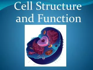
7-2 Eukaryotic Cell Structure • Objectives: • Describe the function of the cell nucleus. • Describe the function of the major cell organelles. • Identify the main roles of the cytoskeleton.
Organelles – Structures that act as specialized organs. Also called “little organs”. Cytoplasm – The portion of the cell outside the nucleus.
Nucleus • Contains nearly all the cell`s DNA and with it the coded instructions for making proteins and other important molecules.
Nucleus Nuclear Envelope – It surrounds the nucleus and is composed of two membranes. It is dotted with nuclear pores, which allows material to move into and out of the nucleus: • Messages • Instructions • Blueprints moving in and out.
Gather your thoughts • What is the nucleulus? • Where is the DNA that a nucleus contains? • Why is DNA important?
Organelles That Store, Clean Up, and Support What are the functions of vacuoles, lysosomes, and the cytoskeleton?
Organelles That Store, Clean Up, and Support What are the functions of vacuoles, lysosomes, and the cytoskeleton? Vacuoles store materials like water, salts, proteins, and carbohydrates. Lysosomes break down lipids, carbohydrates, and proteins into small molecules that can be used by the rest of the cell. They are also involved in breaking down organelles that have outlived their usefulness. The cytoskeleton helps the cell maintain its shape and is also involved in movement.
Vacuoles and Vesicles Many cells contain large, saclike, membrane-enclosed structures called vacuoles that store materials such as water, salts, proteins, and carbohydrates.
Vacuoles and Vesicles In many plant cells, there is a single, large central vacuole filled with liquid. The pressure of the central vacuole in these cells increases their rigidity, making it possible for plants to support heavy structures such as leaves and flowers.
Vacuoles and Vesicles Vacuoles are also found in some unicellular organisms and in some animals. The paramecium contains an organelle called a contractile vacuole. By contracting rhythmically, this specialized vacuole pumps excess water out of the cell.
Vacuoles and Vesicles Nearly all eukaryotic cells contain smaller membrane-enclosed structures called vesicles. Vesicles are used to store and move materials between cell organelles, as well as to and from the cell surface.
Lysosomes Lysosomesare small organelles filled with enzymes that function as the cell’s cleanup crew. Lysosomes perform the vital function of removing “junk” that might otherwise accumulate and clutter up the cell.
Lysosomes One function of lysosomes is the breakdown of lipids, carbohydrates, and proteins into small molecules that can be used by the rest of the cell.
Lysosomes Lysosomes are also involved in breaking down organelles that have outlived their usefulness. Biologists once thought that lysosomes were only found in animal cells, but it is now clear that lysosomes are also found in a few specialized types of plant cells as well.
The Cytoskeleton Eukaryotic cells are given their shape and internal organization by a network of protein filaments known as the cytoskeleton. Certain parts of the cytoskeleton also help to transport materials between different parts of the cell, much like conveyer belts that carry materials from one part of a factory to another. Microfilaments and microtubules are two of the principal protein filaments that make up the cytoskeleton.