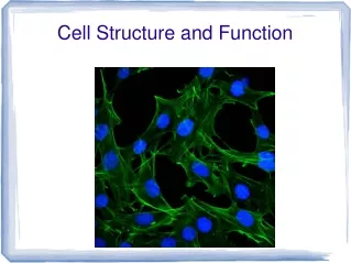
Cell Structure and Function
Cell Structure and Function. Pattern #3: Life Needs an Inside and an Outside. Background Cell Theory Prokaryotes vs. Eukaryotes Surface Area vs. Volume Form vs. Function Cell Membrane Cytoplasma Nucleus. Endoplasmic Reticulum Ribosomes Golgi Apparatus Lysosomes Vacuoles


Cell Structure and Function
E N D
Presentation Transcript
Cell Structure and Function Pattern #3: Life Needs an Inside and an Outside
Background Cell Theory Prokaryotes vs. Eukaryotes Surface Area vs. Volume Form vs. Function Cell Membrane Cytoplasma Nucleus Endoplasmic Reticulum Ribosomes Golgi Apparatus Lysosomes Vacuoles Mitochondria Cytoskeleton Endosymbiosis How Small is SmallUnit Outline
First Glimpse of The Cell • 1665 – Robert Hooke • One of the first microscopists–wrote Micrographia • Thin slices of cork, completely enclosed “cells” • 1677 – Anton Van Leeuwenhoek • First to view living cells – animalcules • Discovered bacteria in the human mouth
Birth of Cell Theory • 1831 – Robert Brown • Discovered nucleus in plant cells • 1838 - Matthias Schleiden • All plants are composed of cells • 1839 – Theodor Schwann • All animals and tissues are composed of cells • 1855 – Robert Remak and Rudolf Virchow • Cells come from other living cells
Cell Theory • All organisms are composed of one or more cells. • Cells are the smallest living things, the basic units of organization for all organisms. • Cells arise only by the division of a previously existing cell.
Prokaryotic Cells • No true nucleus • Still carry out all life functions • Bacteria, cyanobacteria • Cell membrane and sometimes cell wall • Few/no organelles (ribosomes) • Movement by flagella
Eukaryotic Cell • True nucleus • Membrane bound organelles • Compartmentalizaiton
Limits to Cell Size • Communication • Diffusion/Transportation • Surface Area to Volume ratio • Smaller cells have more surface area per unit volume • Larger cells must import/export more materials through the cell membrane • Volume increases at a cubic rate, surface area at a squared rate
The Bigger They Come The Harder to They are to Maintain Cubic Cell Spherical Cell
Some cells are much larger than others. Given the constraints imposed by the S.A. to volume ratio, how would you expect the level of activity in large cells to compare with that in small cells?
Form Follows Function • Nerve cells are long and skinny to transmit messages • Red Blood Cells are close to spherical to maximize S.A. to volume • Skin cells fit together tightly
Just How Small Are We Talking? • Micrometer or micron - m • Nanometer – nm • Angstrom – Å • 1 Å = 0.1 nm = 10-4 m = 10-8 cm • Most cells < 50 m
Plasma MembraneConsistent from Bacteria to Mammals • Forms a protective outer barrier for the cell • Helps maintain a constant internal environment • Regulates exchange of substances in and out of the cell
Fluid-Mosaic Model 1972 – Singer and Nicolson The membrane is made of a phospholipid bilayer that is viscous and free to move. Globular proteins are embedded in the bilayer and move about as well. The Hydrophobic ends of the lipids create a non-polar region within the membrane. This region impedes the passage of all water soluble molecules. Hydrophilic heads exist at the inner and outer surfaces and allow specific chemical interactions to take place.
Membrane Structure • Lipid bilayer • Transmembrane proteins • Network of supporting fibers • Shape and structure • scaffolding • Exterior proteins and glycolipids • “sugar coating” acts as cell identity markers • Glycoproteins – self recognition • Glycolipids – tissue recognition
CytoplasmThe material within a cell excluding the nucleus The cytoplasm of most eukaryotic cells is filled with membranous structures that extend to every nook and cranny of the cell’s interior. While the membranes of the cytoplasm have the same basic structure, the particular proteins embedded in the lipid bilayer vary and give specialized functions. The membranes of the cytoplasm form a highly interdependent network within the cell.
The NucleusRoger, Headquarters • Genetic headquarters • Largest and most easily seen organelle • Repository of genetic information • Discovered by Robert Brown – 1831 • Fungi and other groups may have >1 nucleus • Mammalian erythrocytes (RBC) lose nucleus when mature
Nuclear Structure • Nuclear envelope • Two phospholipid bilayers • Nuclear pores • Membranes pinch together, filled w/ proteins that restrict movement • Proteins moving into the nucleus • RNA and RNA complexes to be exported into the cytoplasm • Nucleolus • Site of intensive rRNA synthesis • Nucleoli • Tiny granules that are precursors to ribosomes • Nucleoplasm • Semifluid area that organizes the contents and provides sites of attachment for enzymes in DNA duplication
Nuclear Shots Nucleus with Pores Nucleus Diagram Nuclear Pore Liver Cell Nucleus
ChromosomesPackaging DNA • Stored as thin strands (chromatin) except for cell division • Allows access to genetic information • During cell division DNA coils around histones in a condensed forms called chromosomes • After cell division chromosomes uncoil and can’t be seen with a light microscope
Endoplasmic ReticulumER every night of the week • Located within the cytoplasm – little net • Highway of the cell • System of passageways that allow materials to be channeled to different locations within the cell • Cell is organized by endomembrane system • ER – lipid bilayer with embedded proteins • Site of membrane phospholipid synthesis • Highly developed in pancreas and salivary glands Smooth ER Rough ER
Do You Like it Rough or Smooth? • Rough ER • Studded with ribosomes • Site of protein synthesis and segregation • Proteins can be used within the cell or exported outside of the cell • Smooth ER • Found in lesser quantities • May be responsible for synthesis of steroids • Break down lipids and toxins in the liver
Golgi Apparatus (Bodies)Delivery System of the Cell • Discovered in 1898 by Camillo Golgi • Flattened stacks of membranes thought to form from vesicles produced by the RER • Abundant in glandular cells – secretions • Collection, packaging, and distribution • Proteins from ER are modified • Short sugar chains are added (glycoproteins and glycolipids) • Two distinct sides • “cis” end accepts protein packages from ER • “trans” end secretes membrane bound vesicles to be expelled from the cell (exocytosis)
exocytosis Secretory Pathway Trans Cisternae Cis
Ribosomes • Site of protein synthesis • They are not membrane bound • Eukaryotic ribosomes slightly larger than prokaryotic • Consists of small and larger subunits • rRNA and about 50 structural proteins • Cluster on the ER to make protein for export • Free ribosomes make proteins for use within the cell • Functional only when attached to mRNA in the cytoplasm • Ribosomal subunits are manufactured in the nucleolus
Lysosomes and Vesicles • Vesicles transport materials in and out of the cell • Exocytosis • Endocytosis – phago- (solid) and pino- (liquid) • Lysosomes – membrane bound digestive vesicles that arise from the golgi bodies • Lysosomes contain a concentrated mix of digestive enzymes • Catalyze breakdown of protein, NA’s, lipids, carbo’s • Recycle old organelles – mitochondria replaced every 10 days • Lysosomes in metabolically inactive eukaryotic cells dissolve cells from the inside out
Vacuoles • Large fluid containing sacs • In plants they may occupy more than 90 percent of the cell’s volume • Bounded by a single membrane • In addition to water the vacuole may contain gases (O2, N2, and/or CO2), acids, salts, sugars, pigments • In plants the vacuole keeps toxins separate from the rest of the cell and maintain internal pressure which aids in the support of the plant
MitochondriaPowerhouse of the Cell • Site of aerobic respiration • Energy released and ATP produced • Inner membrane (cristae) houses the electron transport system • Mitochondria have their own DNA (mDNA) • Cells do not produce new mitochondria during cell division • Mitochondria divide and partition between new cells
Mitochondria(continued) • Could have been a bacteria-like organism incorporated into another cell 1.5 bya • Mitochondria are particularly numerous in muscle cells • All mitochondria of offspring is maternal • Mitochondria of sperm remain outside fertilized egg • mDNA is inherited maternally
Mitochondria Structure Electron Microscope View
CentriolesMicrotubule Assembly Centers • Centrioles – help to assemble microtubules • Some centrioles contain DNA which helps control synthesis of structural proteins • Help assemble spindle fibers which move and align chromosomes during cell divison
Cytoskeleton • Three main types of components • Microtubules – composed of the protein tubulin • Microfilaments – contractile protein actin • Intermediate filaments – variety of proteins • Interconnected to form an elaborate network • Carry out many functions for the cell • Maintain cell shape • Anchor organelles within the cytoplasm • Help in cell movement • Help to organize the internal contents of the cell
Cytoskeleton Fibers • Actin filaments • About 7 nm in diameter • 2 protein chains loosely twined together • Contraction, “pinching”, and cellular extension • Microtubules • About 25 nm in diameter • A ring of 13 protein protofilaments • Cell movement, transport of materials witin the cell • Intermediate filaments • About 8-10 nm in diameter • The most durable element of the cytoskeleton • Structural stability
Cell Movement • Cell motion is tied to the movement of actin filaments, microtubules or both • Actin filaments can form and dissolve very rapidly allowing cells to change shape quickly • In cells treated with drugs that make microfilaments dissolve all cell locomotion stops
Some Crawl, Some Swim • Some cells use a pseudopod (false foot) • Cytoplasmic oozing forces a “foot” out in a certain direction, the cell then drags itself • Some cells use cilia or flagella to swim • Whip-like flagella and shorter cilia both have a 9 + 2 structure of microtubules in eukaryotes • The beating or turning of these structures propels the cell
Endosymbiosis • Proposes that today’s eukaryotic cells evolved by a symbiosis in which one species of prokaryote was engulfed by and lived inside another species of prokaryote • Mitochondria and chloroplasts are thought to be two prime examples of this theory • Double membranes • Both contain circular DNA similar to bacteria • Mitochondria divide by simple fission
Sources Brum, Gilbert D., L. McKane, and G. Karp. 1994. Biology: Exploring Life, 2d ed. New York: Wiley. Raven, Peter H. and G.B. Johnson. 1999. Biology, 5th ed. New York: McGraw-Hill. http://cellsalive.com/ http://gened.emc.maricopa.edu/bio/bio181/BIOBK/BioBookTOC.html http://www.pbrc.hawaii.edu/~kunkel/gallery