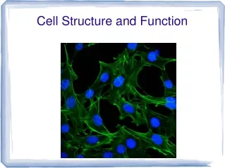
Cell Structure and Function
Cell Structure and Function. Robert Hooke. Robert Hooke. Important Milestones. 1665 – Robert Hooke was first to view and write about cells. He coined the term “cell”. 1674 – Antonie van Leeuwenhoek improved microscopes; was first to see microorganisms.


Cell Structure and Function
E N D
Presentation Transcript
Important Milestones 1665 – Robert Hooke was first to view and write about cells. He coined the term “cell”. 1674 – Antonie van Leeuwenhoek improved microscopes; was first to see microorganisms. 1839 – Schleiden & Schwann claimed that all living tissue (plant & animal) is made of cells. 1858 – Virchow suggested all cells come from other cells, rejecting “spontaneous generation”. The Cell Theory 1. All living things are composed of cells. 2. Cells are the basic units of living organisms. 3. All cells come from pre-existing cells. (Matthias JakobSchleiden, Theodor Schwann, Rudolf Virchow)
onion root intestinal cells amoeba bacteria on pin mitochondrion cell and organelles
Surface area : Volume In a city, too few roads can increase transit time within large blocks; too many roads waste resources and restrict building size. City blocks, agricultural plots, and other artificial boundaries are optimized for transportation Cells similarly transport materials efficiently by optimizing their surface area to volume ratio 5×5×6 150 1×1×750 750 125 5×5×5 5×5×5 125 150÷125 1.2 750÷125 6
All cells have: a membrane to control what enters and leaves, DNA, a source of nutrients & energy, & the ability to eliminate waste. All cells can be classified as prokaryotic or eukaryotic. Prokaryotes and Eukaryotes bacteria animal, plant, protist nucleoid region nucleus (membrane) usually smaller usually larger usually single cell usually multicellular may not need O2 usually needs O2 membrane-bound organelles no organelles
Referred to as “fluid mosaic” model (i.e. lipids and proteins are free to move, & there are diverse elements in varied patterns). extracellular fluid glycoprotein glycolipid Cell membrane cholesterol carbohydrate Phospholipids – self-assemble into a “bilayer” – hydrophilic heads point to watery cytoplasm & extracellular fluid. Hydrophobic tails face each other. Controls what enters and leaves the cell. Cholesterol – helps membrane obtain optimal fluidity (keeps phospholipids apart at low temp., keeps them together at high temp.) peripheral protein filaments of cytoskeleton integral protein cytoplasm
Proteins provide: • structural support by anchoring cytoskeleton • channels for transport • recognition sites to identify the cell (immunity) • receptors, where binding on one side of a protein causes a reaction on the other side In membrane = “integral”, outside = “peripheral” Glycocalyx – polysaccharides extending from the membrane surface, often attached to proteins (glycoproteins). These can help with recognition, lubricate cells, or aid in adhesion. The membrane is more like a soap bubble than a balloon; if punctured it will move, not burst
nuclear pore The Animal Cell chromatin (DNA) nucleus nucleolus nuclear envelope flagellum centriole intermediate filaments cytoplasm rough endoplasmic reticulum plasma membrane ribosome lysosome Golgi apparatus vesicle microtubules smooth endoplasmic reticulum free ribosome mitochondrion vesicle
The Plant Cell Tonoplast Mitochondrion Central vacuole Nuclear envelope Microtubules Nucleus Chromatin Nucleolus Microfilaments Rough endoplasmic reticulum Chloroplast Smooth endoplasmic reticulum Plasmodesmata Peroxisome Ribosomes Cell wall Golgi apparatus Plasma membrane
Animal Cell • cytoplasm • mitochondrion • centriole • lysosome • nucleus • nucleolus • rough endoplasmic reticulum • ribosomes • Golgi apparatus • vesicle smooth endoplasmic reticulum plasma membrane E F D C B A G H I J L K
nucleus • nucleoplasm • nucleolus • nuclear envelope • rough endoplasmic reticulum • smooth endoplasmic reticulum • vacuole • cytoplasm • chloroplast • cell wall Plant Cell • cell membrane • mitochondrion • vesicle • Golgi apparatus
cytosol centrioles nucleolus smooth endoplasmic reticulum nucleoplasm rough endoplasmic reticulum Golgi apparatus vacuoles lysosomes vesicles plasma membrane chloroplast lysosomes nuclear envelope mitochondria cytoskeleton plasma membrane ribosomes cell wall nucleus cytoplasm mitochondria organelles plastids extracellular fluid
B. Plants vs. Animals Only in animals centrioles (plants have centrosome instead) lysosomes Only in plants cell wall chloroplast plastid vacuole (animals have small or no vacuoles)
B. Plants vs. Animals Only in animals centrioles (plants have centrosome instead) lysosomes Only in plants cell wall chloroplast plastid vacuole (animals have small or no vacuoles)