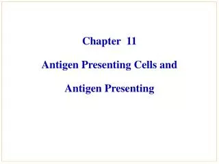
Chapter 11 Antigen Processing and Presentation
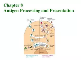
Chapter 11 Antigen Processing and Presentation. B. B. B. B. B. B. B. B. B. Y. Y. Y. Y. Y. Y. Y. Y. Y. Y. T. T. Y. Y. T cells do not recognise native antigens. Y. Y. Y. Y. Y. Y. Cross-linking of surface membrane Ig. Proliferation and antibody production.

Chapter 11 Antigen Processing and Presentation
E N D
Presentation Transcript
B B B B B B B B B Y Y Y Y Y Y Y Y Y Y T T Y Y T cells do not recognise native antigens Y Y Y Y Y Y Cross-linking of surface membrane Ig Proliferation and antibody production No proliferation No cytokine release
T Y Cell surface peptides of Ag presented by cells that express MHC molecules Soluble native Ag Soluble peptides of Ag Cell surface peptides of Ag Cell surface native Ag Antigens must be processed in order to be recognised by T cells APC ANTIGEN PROCESSING T cell response No T cell response No T cell response No T cell response No T cell response
Contents Chapter 11 Antigen Processing and Presentation • PartⅠ Introduction--concepts • PartⅡ Characteristics of APCs • PartⅢ Ag Processing and presentation
PartⅠ Introduction--concepts • Endogenous Ags:antigens synthesized within cells, including self and unself protein----class Ⅰ MHC molecules. • Exogenous Ags: antigens comes outside the cells, including self and unself protein----class Ⅱ MHCmolecules. • Antigen processing:the conversion of native proteins to peptides which can combine with MHC molecules. • Antigen presentation:the course of formation and display of peptide-MHC complexes on the surface of APCsandthe course of peptide-MHC complexes recognition by T cells. • Ag capturing----Endocytosis (internalization) Phagocytosis, Pinocytosis, Receptor-mediated endocytosis
Y The site of pathogen replication or mechanism of antigen uptake determines the antigen processing pathway used EXTRACELLULAR OR ENDOSOMAL REPLICATION Y Vesicular Compartment Contiguous with extracellular fluid Exogenous processing (Streptococcal, tumor antigens) INTRACELLULAR REPLICATION Cytosolic compartment Endogenous processing (Viral, tumor antigens )
T Y Cell surface peptides of Ag presented by cells that express MHC antigens Soluble native Ag Soluble peptides of Ag Cell surface peptides of Ag Cell surface native Ag Antigens must be processed in order to be recognised by T cells ANTIGEN PROCESSING APC APC T cell response No T cell response No T cell response No T cell response No T cell response
Antigen-Presenting Cells (APC) • APC (Accessory cells) : A group of cells play important roles in the immune response which can uptake, process antigens and present peptide-MHC complexes to T cells. • Professional APC: express classⅡMHC molecules Dendritic cell Macrophage B lymphocyte • Facultative APC: endothelial cells, epithelial cells, fibroblast, etc
APC • Express classⅠ, Ⅱ MHC molecules and co-stimulatory molecules • Uptake, process endogenous/exogenous antigens and present peptide-MHC to T cells • Including dendritic cells, macrophages and B cells
PartⅡ Characteristics of APCs • Dendritic cell (DC) • Macrophage • B lymphocyte
1. Dendritic cell (DC) • History:DCs were first found by Steinman in 1973,named for their special spinelike projections. DCs were cultured successfully in vitro in 1993 by Inaba. • Characteristic:The most efficient APC, can present antigens to naive T cells to elicit primary immune response.
1. Dendritic cell (DC) • Identification of DC: Typical morphology—spinelike projection MLR—stimulate naïve T cells activation Surface markers :CD1a, CD11c, CD83(human) high expression of classⅡMHC co-stimulatory molecules--CD80,CD86 others—CKs, CAMs, R (2) Source of DC: pluripotent hematopoietic stem cells myeloid DC myeloid progenitor lymphoid DC lymphoid progenitor GM-CSF, IL-4
1. Dendritic cell (DC) (3) Classification of DC : • DC in lymphoid tissue: Interdigitating DC (IDC) , Folicular DC (FDC) • DC in non lymphoid tissue: Langerhans cell (LC) • DC in body fluid: Veiled cell, Blood DC
Interdigitating DC( IDC ) Express high level of classⅠ, Ⅱ MHC molecules and B7,lack of FcR and CR, can stimulate T cells.
Folicular DC(FDC) FDC Lie in follicle of LN, no expression of class Ⅱ MHC, high level of FcR and C3bR. B cell
Langerhan’s cells(LC)—Birbeck particle Lie in the epithelia of the skin, gastrointestinal and respiratory tracts, express FcR and C3bR. After uptaking antigens, migrating to draining LN and becoming IDC.
1. Dendritic cell (DC) (4) Development and Maturation of DC Pre-DC phase Immature DC(iDC) phase Migration phase Mature DC(mDC) phase
Blood Differentiation Pre-DC Non-lymphoid tissue ImmatureDC Widely distributed in the body Distribute Possess abilityof Ag capture and process Cytokines and Ag DC mature and move into lymphoid tissue Ability of Ag capture and processing decreases while its ability of Ag presenting increases
Difference between iDC and mDC • Ability of uptaking and processing antigens decreases. • Ability of antigen presentation increases. • Express high level of MHC, co-stimulatory molecules(CD80,CD86), CAMs(ICAM-1). • Ability to stimulate naïve T cell activation increases.
1. Dendritic cell (DC) (5) Antigens capturing: • Phagocytosis—cell, bacteria • Pinocytosis—soluble antigen • Receptor-mediated endocytosis FcγRⅡ, C3bR, mannose receptor
(6) Function of DC : Capture, process, present antigens—APC Stimulate T or B lymphocytes—mature DC Induce immune tolerance—immature DC 1. Dendritic cell (DC)
2. Macrophage( MФ) • Stem from monocytes in blood • Have strong phagocytosis (big phagocyte) • Can not stimulate naïve T cells • Capture antigens by phagocytosis, pinocytosis, receptor-mediated endocytosis
Function : Phagocytosis Presentation of antigens Unactivated macrophage Activated macrophage: Class Ⅱ MHC molecules and co-stimulatory molecules 2. Macrophage( MФ)
3. B cells Functions • Mediate humoral immune response • Immunological regulation • Present antigens to T cell Soluble Ag--pinocytosis Specific receptor-mediated endocytosis
T Y Cell surface peptides of Ag presented by cells that express MHC antigens Soluble native Ag Soluble peptides of Ag Cell surface peptides of Ag Cell surface native Ag Antigens must be processed in order to be recognised by T cells ANTIGEN PROCESSING APC APC T cell response No T cell response No T cell response No T cell response No T cell response
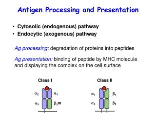
PartⅢ Ag Processing and Presentation • Class Ⅱ MHC pathway ------exogenous antigens • Class Ⅰ MHC pathway ------endogenous antigens • Cross – presentation of antigen
SectionⅠ Class Ⅱ MHCpathway 1. Capture of exogenous Ag 2. Processing of Ag 3. Synthesis and transportation of class Ⅱ MHC molecules 4. Peptide loading of classⅡ MHC molecules 5. Presenting to CD4+T cells
1. Capture of exogenous Ag Endocytosis: Phagocytosis: particles or granules Pinocytosis: soluble antigens Receptor-mediated endocytosis: Form endosome
Y Y Y Uptake of exogenous antigens Membrane Ig receptor mediated uptake Y Phagocytosis Complement receptor mediated phagocytosis Pinocytosis Fc receptor mediated phagocytosis
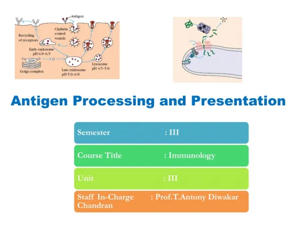
2. Processing of Agendosome + lysosomeAg antigen peptides(10-30aa) Cathepsin
Cell surface Uptake Endosomes To lysosomes Exogenous pathway Protein antigens In endosome Cathepsin B, D and L proteases are activated by the decrease in pH
3. Synthesis and transportation of class Ⅱ MHC molecules Synthesis of class Ⅱ MHC molecules in ER Ii chain --- class Ⅱ MHC molecule(Ii3α3β3 ) ①Promote formation of class Ⅱ MHC dimer ②Preventing endogenous peptide from combining with classⅡMHC molecules within ER ③Leading classⅡMHC molecules into endosome from ER Endosome (MIIC) *Ii chain: Ia-associated invariant chain
and b chains of MHC class II molecules CLIP Invariant chain CLIP peptide A peptide of the invariant chain blocks the MHC molecule binding site. This peptide is called the CLass Ⅱ associated Invariant chain Peptide (CLIP)
MHC class II maturation and invariant chain In the endoplasmic reticulum CLIP Ii chain Invariant chain stabilises MHC class II by non- covalently binding to the immature MHC class II molecule and forming a nonomeric complex Need to prevent newly synthesised, unfolded self proteins from binding to immature MHC
4. Peptide loading of class Ⅱ MHC moleculesIi - class Ⅱ MHC moleculesprotease Ii chain cleavingCLIP - class Ⅱ MHC moleculesHLA-DM CLIP releasingAntigen peptide - class Ⅱ MHC complexes
Cell surface Endosomes Uptake Class II associated invariant chain peptide (CLIP) (inv)3 complexes directed towards endosomes by invariant chain Cathepsin L degrades Invariant chain CLIP blocks groove in MHC molecule MHC Class II containing vesicles fuse with antigen containing vesicles
Removal of CLIP ? How can the peptide stably bind to a floppy binding site? Competition between large number of peptides
HLA-DM HLA-DR Sequence in cytoplasmic tail retains HLA-DM in endosomes HLA-DM catalyses the removal of CLIP HLA-DM Replaces CLIP with a peptide antigen using a catalytic mechanism (i.e. efficient at sub-stoichiometric levels) Discovered using mutant cell lines that failed to present antigen HLA-DO may also play a role in peptide exchange MIIC compartment
5.Presenting to CD4+T cellsAntigen peptide-class Ⅱ MHC molecuels presented on cell membrane by exocytosis
Exported to the cell surface (t1/2 = 50hr) Sent to lysosomes for degradation Surface expression of class II MHC - peptide complexes MIIC compartment sorts peptide-MHC complexes for surface expression or lysosomal degradation