Download
1 / 13
130 likes | 270 Vues
Time Series MRI Core Analysis, Modeling - toward Dynamic Surrogates of Disease. Dominik S. Meier, Ph.D. Center for Neurological Imaging BWH Radiology & Neurology. TSA Paradigm: Capture Processes.

E N D
Time Series MRI CoreAnalysis, Modeling -toward Dynamic Surrogates of Disease Dominik S. Meier, Ph.D. Center for Neurological Imaging BWH Radiology & Neurology
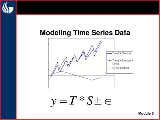
TSA Paradigm: Capture Processes • Research and technology development for longitudinal studies of neurodegenerative disease involving MRI morphometry as outcome measure. • Core work will explore the ability of serial in vivo MRI to illuminate the timing and sequence of the individual pathological processes underlying neurodegenerative disease. • Segmentation of Change vs. Change of Segmentation • Current/Common paradigm: Segmentation -> Trend Analysis • TSA paradigm: Trend Analysis -> Segmentation
Aims • Aim 1: Time Series Fusion • Develop integrated methods for serial image data fusion • concatenates multiple 3-D MRI datasets into a single coherent 4-D space. • spatial and intensity normalization • voxel-based "chronobiopsy" • Aim 2: Time Series Change Detection • Develop a new hierarchical framework for change detection and delineation. • 3-level hierarchy of (1) detection, (2) delineation, and (3) segmentation. • specificity in detection and precision in segmentation • Detection requires high levels of expert knowledge • enhanced precision for delineation requires automation • Aim 3: Time Series Modeling • Develop a framework for change characterization and visualization • parametric models of MRI intensity change • on each voxel time-series profile within the areas of change • investigate the serial MRI data from the viewpoint of a specific biological or clinical hypothesis • "temporal differentiation before spatial integration" • Aim 4: Time Series Validation • Investigate ways to obtain error estimates and sensitivity to change. • scan-rescan data, automated calculation of residual from the fused 4D set • confidence intervals on the model parameters • areas of reference with no pathologic change • sensitivity analyses:sensitivity to change in both the spatial and the intensity domain
Prelim. Results Application: In preparation
T2 intensity R2=0.943 R2=0.943 R2=0.670 0 5 15 20 30 40 50 10 weeks Serial Volumetry Differential Morphometry Segmentation of Change Time Series Modeling Registration Normalization Normalization Spectrum of Serial Morphometry Classifier/Segmentation 2. Classifier/Segmentation 2. TS Modeling 2. Differentiation 3.Classifier / Segmentation 2. Integration 3. Differentiation V(t1) V(t2) V(t3) 3. Differentiation 4. Integration 4. Integration 3.Classifier +Integration Differentiation -> Classification: “new/enlarging” (red),“stable” (green)“resolving” (blue) • Model Required • + Controlled Sensitivity • +Segmentation implied • Greater Expert Input • +segmentation of change • + Controlled Sensitivity - Spatially nonspecific - Sensitive to Registration Error
Technological Biological / Clinical • can dynamic metrics derived from serial MRI provide surrogates with stronger pathological specificity (inflammatory, degenerative, reparatory processes ) ? • Different pathol. processes have different time signatures, even if their morphological footprints remain the same.. • E.g. Inflammation creates mass effect and occurs rapidly. The longitudinalconcept revisited Avoid data reductiondifferentiate first – integrate later • Segmentation of Change vs. Change of Segmentation • Inflammation • Blood Brain Barrier breakdown • Edema • Cellular Infiltration • Degeneration • Demyelination • Axonal Damage • Repair • Macrophage activity • Astrocytosis • Remyelination • Axonal Repair? ~ weeks The cross-sectionalconcept revisited Avoid data reductioncompare first – reduce later • ~ months - years ~ months
t1baseline t2follow-up t3follow-up t4follow-up Data Fusion Pipeline Bias Field Correction coil sensitivity bias Effective spatial resolution loss in serial imaging Partial Volume Filter Registration variable head positioning Segmentation for tissue-specific normalization variable gain, scanner drift, upgrades etc. Intensity Normalization Baseline Normalization Differential: detection of change
Y1: Inflammation / Degeneration Y1 + Y2 Y2: Resorbtion / Repair Two-Process Time Series Model Example: New MS lesion formation We model MRI intensity change as the superposition of two opposing processes, one causing T2 prolongation, another T2 shortening. MRI intensity weeks 0 10 20 30 40 50
T2 intensity MRI intensity complete recovery F1 F3 R2=0.943 F2=0 R2=0.943 partial recovery R2=0.670 F1 F3 F2 weeks 0 5 10 15 20 30 40 50 no recovery F1 F2 F3 weeks Time Series Modeling Example: MS Lesion Formation • F1 = Level of hyperintensity • F2 = Level or recovery • F3 = Duration
Example: Feature Maps of Change F1: Hyperintensity , F2: residual damage , F3: duration [weeks]
Differentiation before Segmentation • sensitivity to change • precision of trend assessment • estimated error in measuringnew lesion change
N=191 N=59 N=43 N=39 100 90 80 70 p=0.25 p=0.003* p=0.33 60 50 40 30 20 10 0 Error Accumulation / Sensitivity Analysis / Pipeline Design Data Modeling Preprocessing Volumetry How one parameter at last step of pipeline affects results is easily tested. The effect of a parameter early in the pipeline is much more difficult to assess. 1 dimension of variation: add and show all results
Conclusions: • Repair does occur in MS, varying in extent by location & subject • MRI intensity dynamics provide reliable metrics of activity • Short-term T2 lesion recovery shows links to progression in both atrophy and disability • SPMSS shows trends to different lesion patterns than RRMS • Dissociation between new lesion size and residual damage“big lesion small damage”, NO equivalence in total lesion burden • Spatial patterns that match histopathological observations