Download
1 / 95
970 likes | 1.03k Vues
Explore the three types of muscle tissue - skeletal, cardiac, and smooth - their functions, and the intricate details of skeletal muscle structure at a microscopic level. Learn about muscle fiber attachments, sarcomeres, sliding filament model of contraction, and the neuromuscular junction.

E N D
Three Types of Muscle Tissue • Skeletal muscle tissue: • Attached to bones and skin • Striated • Voluntary • Powerful
Three Types of Muscle Tissue • Cardiac muscle tissue: • Walls of heart • Striated and contain: • Involuntary
Three Types of Muscle Tissue • Smooth muscle tissue: • In walls of hollow organs (e.g., stomach, urinary bladder, etc.) • Not striated • Involuntary
Muscle Functions • Movement of bones or fluids (e.g., blood) • Maintaining posture and body position • Stabilizing joints • Heat generation (esp. skeletal muscle)
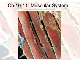
Skeletal Muscle • Connective tissue sheaths of skeletal muscle: • Epimysium: fibrous CT surrounding entire muscle • Perimysium: fibrous CT surrounding fascicles (groups of muscle fibers) • Endomysium: delicate CT surrounding each muscle fiber
Epimysium Epimysium (surrounds entire muscle) Tendon Perimysium Endomysium Perimysium (surrounds a fascicle) Endomysium (surrounds muscle fibers) Muscle fiber
Skeletal Muscle: Attachments • Muscles attach to bone by an origin and insertion • Origin —is fixed and on the immovable bone • Insertion—is on the movable bone • As a contraction occurs the insertion moves towards the origin
Microscopic Anatomy of a Skeletal Muscle Fiber • Sarcolemma = plasma membrane of the muscle fiber (cell) • Multiple peripheral nuclei • Many mitochondria
Microscopic Anatomy of a Skeletal Muscle Fiber • Sarcoplasmic reticulum (SR) • Network of smooth endoplasmic reticulum (SER) surrounding each myofibril • Stores Ca2+ • T (transverse) Tubules • Are continuous with the sarcolemma • Penetrate cell’s interior at each A band–I band junction • T tubules conduct impulses deep into muscle fiber
Sarcolemma Mitochondrion Myofibril Dark A band Light I band Nucleus
Myofibrils • Rod-like structures that fill a muscle fiber (cell) • Myofibrils exhibit striations due to perfectly aligned repeating series of dark A bands and light I bands • Dark A bands and light I bands are part of smaller units called sarcomeres
Sarcomere • Sarcomeres are the smallest contractile unit of a muscle fiber • The Z discs mark the boundaries of a single sarcomere • Sarcomeres are composed of thick and thin filaments
Regions of a Sarcomere • A band (Dark Band)- Contains thin & thick filaments • H zone: lighter midregion where filaments do not overlap • M line: proteins that hold adjacent thick filaments together; center of sarcomere • I band (Light Band)- Contains only thin filaments • Z disc: marks the start and end of one sarcomere
Thin (actin) filament Z disc H zone Z disc Thick (myosin) filament I band A band Sarcomere I band M line (c) Part of one myofibril Sarcomere Z disc Z disc M line Thin (actin) filament Elastic (titin) filaments Thick (myosin) filament (d)
Structure of a Thick Filament • Thick filaments • are composed of many myosin proteins • A single myosin protein has a head and tail region • The myosin head binds actin and pulls it during a contraction • The myosin head breaks down ATP to release energy for a muscle contraction
Structure of a Thin Filament • Thin filaments (actin) • are twisted strands of actin protein • have binding sites for the myosin head
Longitudinal section of filaments within one sarcomere of a myofibril Thick filament Thin filament Thick filament Thin filament Portion of a thick filament Portion of a thin filament Myosin head Tropomyosin Troponin Actin Actin-binding sites Active sites for myosin attachment Tail Heads Actin subunits ATP- binding site Flexible hinge region Myosin molecule Actin subunits
I band A band I band Z disc H zone Z disc Myofibril M line Sarcolemma A Triad: 1 T tubule • • 2 terminal cisternae of the SR SR Myofibrils Mitochondria
Sliding Filament Model of Contraction • In the relaxed state, thin and thick filaments slightly overlap • During contraction, myosin heads bind to actin, detach, and bind again, pulling thin filaments toward M line • As H zones shorten and disappear, sarcomeres shorten, muscle cells shorten, and the whole muscle shortens
Z Z H A I I 1 Fully relaxed Z Z I A I 2 Fully contracted
The Neuromuscular Junction • Axons of motor neurons travel from the brain/spinal cord via nerves to skeletal muscles • Each axon branches into a number of axon terminals as it enters a muscle • Each axon ending forms a neuromuscular junction with a single muscle fiber
Myelinated axon of motor neuron Action potential (AP) Axon terminal of neuromuscular junction Nucleus Sarcolemma of the muscle fiber 1 Action potential arrives at axon terminal of motor neuron. Ca2+ Synaptic vesicle containing ACh Ca2+ 2 Voltage-gated Ca2+ channels open and Ca2+ enters the axon terminal. Mitochondrion Synaptic cleft Axon terminal of motor neuron Fusing synaptic vesicles Figure 9.8
The Neuromuscular Junction • An axon terminal and muscle fiber are separated by a space called the synaptic cleft • Synaptic vesicles within axon terminal contain the neurotransmitter acetylcholine (ACh) • Junctional folds of the sarcolemma contain ACh receptors
Events at the Neuromuscular Junction • A nerve impulse arrives at the axon terminal of a motor neuron • Ca2+ floods into axon terminal • Ca2+ entry to the axon terminal causes the release of Ach • ACh diffuses across synaptic cleft and binds to receptors on the sarcolemma • ACh binding opens sodium channels • Na+ floods into muscle fiber and K+ floods out making the interior of cell less negative (depolarization) • Once a threshold is reached an AP is generated
Axon terminal Open Na+ Channel Closed K+ Channel Na+ Synaptic cleft ACh K+ Na+ K+ + + + + ACh + + + + + + Action potential n + + o i t Na+ K+ a z 2 i r Generation and propagation of the action potential (AP) a l o p e d f o e v a W 1 1 Local depolarization: generation of the end plate potential on the sarcolemma Sarcoplasm of muscle fiber
The Action Potential • The AP is an unstoppable, electrical event that travels along the entire sarcolemma conducting the electrical impulse from one end of the cell to the other • The result of the AP is a muscle contraction
Setting the stage Axon terminal of motor neuron Action potential is generated Synaptic cleft ACh Sarcolemma Terminal cisterna of SR Ca2+ Muscle fiber Triad One sarcomere
Destruction of Acetylcholine • ACh effects are quickly terminated by the enzyme acetylcholinesterase (AChE) • AChE prevents continued muscle fiber contraction in the absence of additional stimulation by a motor neuron
1 Action potential spreads along the sarcolemma and down the T tubules. Steps in E-C Coupling: Sarcolemma Voltage-sensitive tubule protein T tubule Ca2+ release channel 2 Calcium ions are released from the SR. Terminal cisterna of SR Ca2+
Actin Binding sites on actin are blocked Ca2+ Myosin 3 Calcium unblocks binding sites on actin Active sites exposed and ready for myosin binding Myosin binds actin and the contraction begins 4 Myosin cross bridge The aftermath
Role of Calcium (Ca2+) in Contraction • At low intracellular Ca2+ concentration: • Binding sites on actin are blocked • Myosin heads cannot attach to actin, so no contraction (muscle fiber is relaxed)
Role of Calcium (Ca2+) in Contraction • At higher intracellular Ca2+ concentrations: • Ca2+ causes binding sites on actin to be exposed • Myosin binds actin (cross bridge cycle occurs) • When nervous stimulation ceases, Ca2+ is pumped back into the SR and contraction ends
Cross Bridge Cycle • Continues as long as the Ca2+ signal and adequate ATP are present • Cross bridge formation: high-energy myosin head attaches to thin filament • Power stroke: myosin head pivots and pulls thin filament toward M line
Cross Bridge Cycle • Cross bridge detachment: ATP attaches to myosin head and the cross bridge detaches • “Cocking” of the myosin head: energy from breaking down ATP cocks the myosin head into the high-energy state
Thin filament Ca2+ Actin ADP Myosin cross bridge Pi Thick filament Myosin Cross bridge formation. 1 ADP ADP Pi ATP hydrolysis Pi The power (working) stroke. 4 2 Cocking of myosin head. ATP ATP Cross bridge detachment. 3
Motor Unit: The Nerve-Muscle Functional Unit • A motor unit includes a motor neuron and all muscle fibers it supplies (can be four to several hundred)
Spinal cord Axon terminals at neuromuscular junctions Motor unit 1 Motor unit 2 Nerve Motor neuron cell body Motor neuron axon Muscle Muscle fibers Figure 9.13a
Graded Muscle Responses • Defined: Variations in the degree of muscle contraction Responses are graded by: • Changing the frequency of stimulation • Changing the number of muscle cells being stimulated at one time (by changing strength of stimulus)
Response to Change in Stimulus Frequency • A single stimulus results in a single contractile response called a muscle twitch
Single stimulus single twitch Contraction Relaxation Stimulus A single stimulus is delivered. The muscle contracts and relaxes Figure 9.15a
Response to Change in Stimulus Frequency • Increase frequency of stimulus muscle doesn’t have time to completely relax (btwn. stimuli) • Ca2+ release stimulates further contraction temporal (wave) summation • Further increase in stimulus frequency unfused (incomplete) tetanus
Low stimulation frequency unfused (incomplete) tetanus Partial relaxation Stimuli (b) If another stimulus is applied before the muscle relaxes completely, then more tension results. Figure 9.15b
Response to Change in Stimulus Frequency • If stimuli are given quickly enough, fused (complete) tetanus results
High stimulation frequency fused (complete) tetanus Stimuli (c) At higher stimulus frequencies, there is no relaxation at all between stimuli. This is fused (complete) tetanus. Figure 9.15c
Muscle Metabolism: Energy for Contraction • ATP is the only source used directly for contractile activities • Available stores of ATP are depleted in 4–6 seconds
Muscle Metabolism: Energy for Contraction • ATP is regenerated by: • Direct phosphorylation of ADP by creatine phosphate (CP) • Anaerobic pathway • Aerobic pathway
Muscle Metabolism: Energy for Contraction • Direct phosphorylation of ADP by creatine phosphate (CP) • CP is more concentrated in muscle fibers than ATP (~4 X more) • When ATP stores are depleted: muscle fibers use CP to regenerate ATP • Products are: 1 ATP/ CP • Provides energy for: ~ 15 seconds of activity
(a) Direct phosphorylation Coupled reaction of creatine phosphate (CP) and ADP Energy source: CP CP ADP Creatine kinase Creatine ATP Oxygen use: None Products: 1 ATP per CP, creatine Duration of energy provision: 15 seconds Figure 9.19a