Chapter 6 - Integumentary Sx.
Chapter 6 - Integumentary Sx. The Skin and Accessory Structures. Integumentary Sx.


Chapter 6 - Integumentary Sx.
E N D
Presentation Transcript
Chapter 6 - Integumentary Sx. The Skin and Accessory Structures
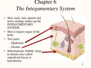
Integumentary Sx. • The skin is one of the largest organs in the body and serves many functions. Chief among them is protection. It also prevents water loss and regulates body temperature. It also acts as a general sense organ for the sense of touch and tactile input.
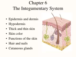
Integumentary Sx. • The skin has three layers: • Epidermis – composed primarily of stratified squamous epithelium. • Dermis – thicker than the epidermis, this layer is composed of connective tissue, smooth muscle, nervous tissue and blood vessels. • Hypodermis – aka subcutaneous layer, the hypodermis is composed of areolar tissue and adipose.
Integumentary Sx. The Epidermis • The lowest level of cells in this layer is the foundation of the entire layer. This layer of cells is called stratum basale and sits atop the basement membrane. These cells are closest to the underlying blood supply in the area and thus receive the best nourishment. These cells are almost always undergoing cell division and new cells are pushed up and away from the blood supply. This rise in successive levels removes the cells farther from the blood supply and in time they die.
Integumentary Sx. • As these epithelial cells rise in layers many of them undergo the process of keratinization. During this process the cytoplasm of the cell is replaced by a fibrous waterproofing protein called Keratin.
Integumentary Sx. • The successive layers of the epidermis are as follows: • Stratum Basale – bottom most layer, mitosis is ongoing • Stratum Spinosum • Stratum Granulosum – keratinization is taking place, these cells are dying • Stratum Lucidum • Stratum Corneum – completely keratinized, these cells are all dead and ready to be shed.
Integumentary Sx. • Calluses are a thickening of the epidermis due to regular friction or pressure. • Melanocytes are specialized cells in the epidermis that reside in the stratum basale. They produce a pigment called melanin that provides skin color. • Melanocytes are stimulated by ultraviolet radiation to produce melanin that absorbs some of that form of radiation.
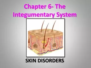
Integumentary Sx. Skin Cancer **Skin cancer is the most common form of cancer and usually poses the least threat to life and health. If caught early it is usually very easily treated and exhibits very little incidence of return. • Basal Cell Carcinoma – the most common form of skin cancer, this lesion is benign and very seldom metastasizes (spreads) to distant sites. • Squamous Cell Carcinoma – the next most common form of skin cancer, this carcinoma is almost as reluctant to metastasize as the previous mentioned condition. • Malignant Melanoma – making up only a very small minority of skin cancers (about 5% or less), but is one of the most dangerous forms of any cancer known. Caught early it is similar in treatment to the above forms. However, metastasis of melanoma can be very fast, and once this happens the survival rate after treatment plummets greatly.
Integumentary Sx. The Dermis • The Dermis is largely composed of connective tissues with a lot of collagen and elastic fibers. The dermis has many accessory structures of the skin located in it. • Nerve endings scattered throughout the dermis are responsible for the variety of sensation available thru the skin. • The papillary layer of the dermis is responsible for fingerprint patterns.
Integumentary Sx. Accessory Structures • Hair Follicles: Hair grows from epidermal cells found in the dermis. Growth of hair starts at the root where blood vessels nourish the hair. The shaft of hair is made up of keratinized epithelium. • Hair color is determined by genetics. The more melanin a person produces, the darker their hair is.
Integumentary Sx. • A bundle of smooth muscle cells called arrector pili is attached to each hair follicle. This muscle is responsible for making the hair stand up at times and thus for goose bumps. Arrector pili are under autonomic control. • Nails: these structures are made of a special keratinized epithelium. Each nail has a free edge, nail plate, nail bed, a lunula and a root or matrix.
Integumentary Sx. • Skin Glands: the glands in the dermis are considered exocrine glands as they possess a duct for secretory purposes. • Sebaceous Glands – these glands secrete body oil known as sebum. These glands are usually associated with a hair follicle. Sebum helps the body by keeping the skin and hair soft, pliable and waterproof. An underproduction will result in dry skin and brittle hair.
Integumentary Sx. • Sudoiferous Glands – There are two types of sweat glands: • Eccrine – these are the smallest and most numerous sweat glands. They are found body wide with few exceptions. • Apocrine – larger than eccrine glands, these glands also secrete sweat. Found only in the axilla, groin, and around the nipples, apocrine glands do not become active until puberty. They usually have a small amount of melanin associated with them, thus areas containing these glands are slightly darker in color.
Integumentary Sx. Regulation of Body Temperature • Normal body temperature ranges from 96.5-99.5 degrees F. Average body temperature is 98.6 degrees F. or 37 degrees C. • Heat in the human body is a product of metabolic reactions and muscle contractions.
Integumentary Sx. • Heat is lost from the body in a number of ways: • Radiation – loss of heat in the form of infared energy from a warm surface to cooler surroundings. • Conduction – loss of heat from the body into an object that is in direct contact with the body.
Integumentary Sx. • Convection – The loss of heat from the body due to the movement of cooler air over the body. • Evaporation – the loss of heat from the body by way of moisture as it leaves the surface of the body as a gas. • The body may retain or release heat thru the action of the blood vessels. By constricting the diameter of the superficial vessels the warmth of the blood is conserved in the body’s core areas such as the torso and head. Pallor and perhaps cyanosis can result in this situation. On the other hand, if the blood vessels are dilated, then more blood reaches the skin and is loss by means of radiation and flushing will probably result.
Integumentary Sx. • Hypothermia is a condition where the core body temperature falls below a manageable level. A gradualre-warming of the body is called for in order to restore homeostasis. • Hyperthermia is a condition where the core body temperature rises above a manageable level. Two types are noted: • Heat Exhaustion – a condition that may be easily managed by cooling of the body as well as the replacement of body fluids and needed electrolytes. • Heat Stroke – a condition that is considered a medical emergency as it poses a threat to life. The tx. is the same as above, but will likely need to be done in a medical setting as the fluids are often administered via IV therapy.
Integumentary Sx. • Skin color is due to a variety of factors. Included are genetics (heritage), environmental factors (sun exposure or lack of), and physiological factors (such as flushing and cyanosis). • The three pigments playing into skin color are melanin, carotene (found in certain foods), and hemoglobin (found in red blood cells). • Albinism is a condition where a person’s body does not make any of the melanin pigment.
Integumentary Sx. • Inflammation is a response of tissue to injury. The five signs of inflammation are: • Reddening • Swelling • Heat • Pain • Loss of Function
Integumentary Sx. Burns • Burns may be classified into catagories by the depth to which they penetrate the body: • First degree burns are also called superficial partial thickness burns. They are characterized by a slight reddening of the skin. The damage does not pass beyond the epidermis and usually heals within a few days. • Second degree burns are also called a deep partial thickness burn. These are characterized by blisters. Second degree burns penetrate thru the epidermis into the dermis but no farther. Blistering is due to damaged capillary vessels. These burns are usually quiet painful. Healing time will usually be at least two weeks, but may take longer depending on how extensive the damage to the skin.
Integumentary Sx. • Third degree burns are also called full thickness burns. These burns penetrate thru the epidermis, thru the dermis, into the hypodermis and possibly much deeper. Charring of the tissues is evident in a third degree burn, but the are of the third degree burn is without pain as the nerve endings in the area are destroyed. Healing time can be quite extensive and surgical grafts are frequently used to assis in the process.
Integumentary Sx. • Burn patients are assessed using the rule of nines. This is a system by which the body is assigned an amount of surface are based on size. See chart p.187.