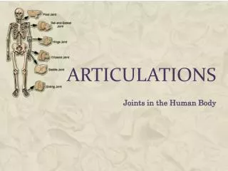
Chapter 9: Articulations
Chapter 9: Articulations. Articulations. Body movement occurs at joints where 2 bones connect Articulation = joints; site where two or more bones meet. Joint Structure. Determines direction and distance of movement ( range of motion ) Joint strength decreases as mobility increases.

Chapter 9: Articulations
E N D
Presentation Transcript
Articulations • Body movement occurs at joints where 2 bones connect • Articulation = joints; site where two or more bones meet
Joint Structure • Determines direction and distance of movement (range of motion) • Joint strength decreases as mobility increases
Anatomical/Structural Classification of Joints (based on connecting material • Fibrous: bones joined by fibrous CT with no space • Cartilaginous: bones joined by pad or bridge of cartilage • Synovial: bones separated by fluid-filled cavity, surrounded by CT
Structural Classification Table 9–2
Physiological/Functional Classifications of Joints:(based on the amount of movement) • Synarthrosis: immovable joints - No movement - Fibrous or cartilaginous connections • Amphiarthrosis: slightly moveable joint - Little movement - Fibrous or cartilaginous connections • Diarthrosis: freely moveable joint - More movement - always synovial connections
What common characteristics do typical synarthrotic and amphiarthrotic joints share? joint capsules filled with fluids non-restricted movement of bony regions bony regions separated by fibrous connective tissue articular cartilages and bursae
Synarthroses (Immovable Joints) • Are very strong • Edges of bones may touch or interlock • 4 Types of Synarthrotic Joints • Suture • Gomphosis • Synchondrosis • Synostosis
1. Synarthoroses * Immoveable Strength • Synotosis: fused bones - Suture of skull and epiphyseal lines of long bones • Suture: interlocked bones, sealed with dense CT - Found only in the skull • Gomphosis: tooth in alveolar socket, held by peridontal ligament - Binds teeth to sockets • Synchrondrosis: hyaline cartilage bridge between bones - Epiphyseal cartilage of long bones and between vertebrosternal ribs and sternum
2. Amphiarthroses * Slightly Moveable, strength with some mobility • Syndesmosis: bones connected by ligament • Symphasis: bones separated by pad of fibrocartilage
3. Diarthorses = Synovial Joints • Great mobility, less strength and stability • At ends of long bones • Within articular capsules • Lined with synovial membrane
Features of Synovial Joints • Articular Cartilage • Synovial Cavity • Articular Capsule • Synovial Fluid • Accessory Structures - Meniscus, fat pad, accessory ligaments, tendons, bursa, synovial tendon sheath
Marrow cavity Spongy bone Periosteum Synovial membrane Articular cartilage Joint cavity (containing synovial fluid) Articular capsule Compact bone (a) Quadriceps tendon Bursa Joint capsule Femur Patella Synovial ligament Articular cartilage Fat pad Meniscus Patellar ligament T ibia Joint cavity Meniscus Intracapsular ligament (b)
1. Articular Cartilages • Hyaline cartilage • No perichondrium or periosteum • Pad articulating surfaces within articular capsules: • prevent bones from touching • Smooth surfaces lubricated by synovial fluid: • reduce friction
Features of Synovial Joints • Synovial cavity: • Space between/around opposing bones • Has synovial fluid • Articular Capsule: 2 layers • Outer • dense irregular connective tissue, continuous with periosteum • Inner • synovial membrane (areolar CT), covers inside surface of cavity except articular cartilage, secretes synovial fluid
4. Synovial Fluid • filtrate from blood plasma + hyaluronic acid from fibroblasts * Functions: 1. Lubrication 2. Nutrient distribution: diffusion medium 3. Shock absorption
Synovial Joints: Accessory Structures • Cartilages (Meniscus) • Fat pads • Ligaments • Tendons • Bursae • Synovial Tendon Sheath
Synovial Joints: Accessory Structures • Meniscus: • fibrocartilage pad = articular disc • Subdivides cavity or changes shape of articular surface, limits range of motion • Cushion the joint • Fat Pad: • Adipose • Superficial to joint capsule, • Function: protection and space filler
Synovial Joints: Accessory Structures • Accessory Ligaments: • Dense regular connective tissue • Either part of capsule, inside joint, or outside capsule • Function: support and strengthen joint • Sprain: Ligaments with torn collagen fibers • Tendons: • Dense regular connective tissue • Attach muscle to bone • Function: Add stability to the joint
Synovial Joints: Accessory Structures • Bursa: • Synovial fluid filled pocket • Function: reduces friction • Cushion areas where tendons or ligaments rub • Synovial Tendon Sheath: - Tubular bursa around a tendon
Synovial Joints: Stabilizing Factors • Prevent injury by limiting range of motion: • collagen fibers (joint capsule, ligaments) • articulating surfaces and menisci • other bones, muscles, or fat pads • tendons of articulating bones
Functional Classification Table 9–1
In a newborn infant, the large bones of the skull are joined by fibrous connective tissue. Which type of joints are these? The bones later grow, interlock, and form immovable joints. Which type of joints are these? synarthrosis; gomphosis symphysis; sutural synchondrosis; synostosis syndesmosis; sutural
Why would improper circulation of synovial fluid lead to the degeneration of articular cartilages in the affected joint? Synovial fluid nourishes articular cartilage. Blood flow follows synovial fluid circulation. Articular cartilage is composed of synovial fluid. Both A and B.
Joint Injuries • Sprain: damage to ligament • Some collagen torn, slow to heal • Bursitis: • Inflammation of a bursa due to trauma, infection, or repetitive motion ** Synovial joints stabilized by articular capsule and accessory structures to restrict mobility: Increase Mobility = Decrease Stability = Increase chance of dislocation
Injuries • Luxation: • Dislocation • articulating surfaces forced out of position • Joint Displacement • Usually damages articular cartilage, ligaments, and joint capsule • Pain receptors in all CT of the Joint, except articular cartilage, to prevent actions • Subluxation: • Partial dislocation • Displacement beyond usual anatomical limitation • “Double Jointed”
Movements at Synovial Joints • Linear Movements: • Gliding: slight movement in any direction • Angular Movements: one plane of motion • Flexion: reduce angle in frontal plane • Extension: increase angle in frontal place • Hyperextension: extension past anatomical position • Abduction: move away from longitudinal axis in sagittal plane • Adduction: move toward longitudinal axis in sagittal plane • Circumduction: move in loop without rotation • Rotational Movements: turn on axis • Medial Rotation: turn in toward body • Lateral Rotation: turn out away from body
Linear Motion • Pencil maintains vertical orientation, but changes position Figure 9–2a, b
Angular Motion • Pencil maintains position, but changes orientation Figure 9–2c
Circumduction • Circular angular motion Figure 9–2d
Rotation • Pencil maintains position and orientation, but spins Figure 9–2e
Angular Motion: Flexion Figure 9–3a
Angular Movement: Flexion Vs. Extension • Flexion • Angular motion • Anterior–posterior plane • Reduces angle between elements • Extension • Angular motion • Anterior–posterior plane • Increases angle between elements
Angular Motion: Abduction Vs. Adduction Figure 9–3b, c
Abduction Vs. Adduction • Abduction: • Angular motion • Frontal plane • Moves away from longitudinal axis • Adduction: • Angular motion • Frontal plane • Moves toward longitudinal axis
Circumduction • Circular motion without rotation • Angular motion Figure 9–3d
Special and Specific Motion • Inversion: turn sole inward • Eversion: turn sole outward • Dorsiflexion: lift toes • Plantar flexion: lift heal • Opposition: thumb across palm • Pronation: medial rotation of radius • Superination: lateral rotation of radius • Protraction: move anterior (toward front) • Retraction: move posterior (toward back) • Elevation: move superior (toward head) • Depression: move inferior (toward feet)
Inversion and Eversion Figure 9–5a
Dorsiflexion and Plantar Flexion Figure 9–5b
Ranges of Motion • Monaxial: movement in 1 plane • Biaxial: movement in 2 planes • Triaxial: movement in 3 planes • Multiaxial: gliding joints, all directions
When you do jumping jacks, which lower limb movements are necessary? flexion and extension abduction and adduction flexion and abduction plantar flexion and eversion
The types of synovial joints, and the relationship of motion to structure.
Classification of Synovial Joints by Shape • Gliding • Hinge • Pivot • Ellipsoidal • Saddle • Ball-and-socket
Gliding/Plane Joints • Flattened or slightly curved faces • Slide in any direction Figure 9–6 (1 of 6)
Hinge Joints • Cylindrical projections in trough-shaped surface • Angular motion in a single plane (monaxial) Figure 9–6 (2 of 6)
Pivot Joints • Round projection in ring shaped depression • Rotation only (monaxial) Figure 9–6 (3 of 6)
Ellipsoidal Joints • Oval articular facet within an oval depression • Motion in 2 planes (biaxial) Figure 9–6 (4 of 6)
Saddle Joints • 2 concave faces into convex • Straddled (biaxial) Figure 9–6 (5 of 6)
Ball-and-Socket Joints • Spherical head into cup-like socket • Round articular face in a depression (triaxial) Figure 9–6 (6 of 6)