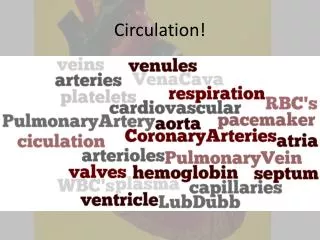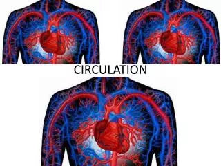
Understanding the Human Circulatory System: Functions and Components
The human circulatory system plays a vital role in transporting blood throughout the body, consisting of approximately 96,000 km of blood vessels. It pumps around 5 liters of blood every minute, delivering oxygen and nutrients to cells while removing waste products. The system includes arteries, arterioles, capillaries, venules, and veins, each with specific functions. Key elements like the heart, atria, ventricles, and valves ensure proper blood flow direction. Understanding this system is essential for recognizing its importance in health and disease.

Understanding the Human Circulatory System: Functions and Components
E N D
Presentation Transcript
Circulation The human circulatory system consists of 96,000 km of blood vessels that transport blood to each cell in the body. Your entire blood volume (about 5L) is pumped every minute.
The circulatory system performs the following functions: 1. carries oxygen and nutrients to the cells 2. carries carbon dioxide and waste away from the cells 3. carries hormones to target organs 4. distributes heat throughout the body 5. helps defense of invading micro-organisms
Blood Vessels • Arteries – carry blood away from the heart. Their thick walls are composed of three distinct, elastic layers. Each time the heart pumps, the arteries stretch to accommodate the rush of blood. This is felt in the neck, or on the wrist as a pulse. • Arterioles - are smaller arteries whose middle layer is composed of elastic fibers and smooth muscle. The arterioles are able to contract and relax, controlling blood flow to different parts of the body.
Vasoconstriction – the narrowing of blood vessels, decreasing flow to the tissues. Vasodilation – the widening / relaxation of blood vessels, increasing flow to the tissues.
Capillaries – are tiny blood vessels composed of a single layer of cells. This is the site of fluid and gas exchange between the cells and the body tissues. Many capillaries are only as thick in diameter as one red blood cell (<0.005 mm). Pressure in the capillaries is high, increasing the risk of rupturing and causing a bruise.
Venules – larger blood vessels that form as capillaries merge. The venules are lined with smooth muscle to ensure blood continues to flow back towards the heart. • Veins – larger blood vessels that result as venules merge, take blood back towards the heart. Veins also serve as blood reservoirs, holding up to 65% of the total blood volume. Blood pressure in the veins is quite low, so the veins have uni-directional valves that ensure the one way flow of blood. Skeletal muscles also help aid venous flow. Venous pressure increases when skeletal muscles contract and push on the vein, forcing blood upwards.
Problems with blood vessels: • Aneurysm – a bulge or weakening in the wall of a blood vessel. • Atherosclerosis – degeneration of blood vessels caused by the accumulation of fat deposits (plaque) in the inner wall.
Bruising – rupture of capillary beds cause blood to leak into the extra-cellular space. • Varicose Veins – damage to the one-way valves in veins causes blood to pool and the veins to bulge.
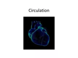
Circulation • The heart consists of two parallel pumps. The right connects to blood vessels that circulate blood to the lungs, for oxygenation, and back to the heart. This system is called the pulmonary circulatory system. The second, left hand pump, connects blood vessels to the body and circulates blood to the body tissues. This system is called the systemic circulatory system. One way blood flow is maintained by uni-directional valves in the heart and in the blood vessels.
Pulmonary System Systemic System
Atria - top chamber of the heart that contracts to push blood into the bottom ventricular chamber. • Aorta – largest artery in the body, carries oxygenated blood to the body tissues. • Atrioventricular (AV) valves – the tricuspid (Right side) and bicuspid valves (left) separate the atria from the ventricles on the right and left sides respectively. These valves ensure a one way flow of blood within the heart. Chordae tenidea anchor the valves to ensure one way flow. • Coronary arteries – branch from the aorta and supply the heart muscle with oxygenated blood.
Pulmonary arteries / veins – carry deoxygenated blood to (artery) and oxygenated blood from (vein) the lungs. • Semi-lunar valve – prevents blood from flowing backwards from the pulmonary artery / vein, back to the ventricles. • Septum – muscular wall that separates the left and right sides of the heart. • Vena Cava – largest vein that carries blood from the upper body (superior) or lower body (inferior) back to the heart. • Ventricles – large muscular chambers that pump blood to the lungs (right), or to the body (left). The wall of the left ventricle is thicker because it pumps blood further to the body tissues.
Blood Flow Through The Heart • Blood always flows in one direction through the heart: • Body cells (deoxygenated • Inferior / Superior Vena Cava • Right Atrium • Right Ventricle • Pulmonary Artery (to lungs) • Pulmonary Vein (from lungs) • Left Atrium • Left Ventricle • Aorta • HeartCenterOnline: • Blood Flow Through Your Heart and Lungs
Setting the Heart Beat • The heart muscle is unique because it is myogenic muscle. That is, it is able to contract without external nerve stimulation. The heart beat is controlled by the sympathetic (stimulating) and parasympathetic (relaxing) branches of the nervous system. The heart’s rate or tempo is set by a small mass of tissue in the right atrium called the sinoatrial (SA) node. This is known as the muscles pacemaker. Contractions radiate from the SA node and travel to the atrioventricular node (AV) node through the bundle of His, which passes nerve impulses along two large nerve fibers called Purkinje fibers to the septum and the ventricles. The resulting wave of contraction moves from the atria to the ventricles, up from the bottom of the ventricles, forcing blood out the atria / pulmonary arteries. • http://www.msjensen.gen.umn.edu/1135/Links/Animations/Flash/0028-swf_the_cardiac_cy.swf
Normal Heart Rate – 80 beats/minute Bradycardia - < 50 beats/min Tachycardia - > 100 beats/min Systole – contraction of the heart muscle Diastole – relaxation of the heart muscle Electrical Signals and Blood Flow
ECG • ElectroCardioGram • Shows the electrical conductivity of the heart and is used to identify and diagnose heart conditions. • Heart Institute: Heartbeat Animation
Heart Sounds and Cardiac Cycle • The familiar lubb-dubb sound of the heartbeat is caused by the closing of the heart valves.
Heart Murmur (lubb-dubb-squish)– occurs when the heart valves are faulty and don’t close completely. Blood rushes from the ventricle back into the atrium creating a squishing sound that is heard as a murmur. • Transport: Cardiovascular System - Heart Sounds
Cardiac Output and Blood Pressure • Cardiac Output – is the amount of blood that flows out of the heart each minute. Output can be influenced by stroke volume and heart rate. • Stroke volume – is the volume of blood pumped with each beat of the heart. On average, the stroke volume is about 70 ml/beat. • Heart rate – is the number of beats per minute of the heart. Cardiac Output = Stroke volume x Heart rate
Example: • If Tom has a stroke volume of 85 mL and a heart rate of 100 beats per minute, what is his cardiac output? If the dog has a stroke volume of 50 ml and a heart rate of 120 beats per minute, what is his cardiac output? • Tom: 85 mL/beat x 100 beats/min = 8500mL/min. • Dog: 50 mL/beat x 120 beats/min = 6000 mL/min.
Blood Pressure – is the force of the blood on the walls of your arteries. Blood pressure is measured with a stethoscope and a sphygmomanometer (or blood pressure cuff). Normal blood pressure is 120/80. That means that the systolic pressure (during contraction) is 120 mm Hg and the diastolic pressure (during relaxation) is 80 mm Hg. Blood pressure is influenced by age, level of fitness, level of hydration and level of stress.
Blood pressure depends on two major factors: BP is measured by baroreceptors (stretch receptors) in your aortic arc or the carotid artery • 1. Cardiac output (blood volume and Heart Rate) – higher blood volume = higher pressure, this may also be influenced by fluid retention (such as water). Higher heart rate = higher BP • 2. Arteriolar resistance(blood vessel size)– the diameter of the arteries will determine the pressure within them. Vasoconstriction will cause greater BP. Vasodilatation decreases BP • Ex. High BP will be interpreted by the baroreceptors and signal the medulla omblongta to do 2 things to drop the BP to normal: • 1) decrease the sympathetic nervous system cause vasodilatation • 2) increase the parasympathetic nervous system slow the heart rate and decrease the blood volume
Measuring Blood Pressure A sphygmomanometer (blood pressure cuff)measures blood pressure: Systolic / Diastolic pressure is on average 120/80 mm Hg. Blood Pressure Center - HeartCenterOnline:
Capillary Fluid Exchange • Nearly every tissue of the body is within 0.1 mm of a capillary. The capillaries carry valuable oxygen, glucose and amino acids to cells, as well as remove wastes like carbon dioxide. • Extra-cellular Fluid (ECF) – fluid that occupies the spaces between cells. This fluid will then come into contact with both cells and the capillaries to facilitate the transport of oxygen, glucose, amino acid and waste. • Fluid Pressure/Filtration: BP created by the heart and blood volume filter oxygen and nutrients out of the capillaries at the arteriole end • Osmotic Pressure/Absorption: Pressure created by water pushed CO2 and waste back into the capillaries at the venule end
Arteriole End -A high BP on the inside of the vessel vs. a low osmotic pressure on the outside of the vessel allows for oxygen and nutrients to be squished outwards and into the ECF to be picked up my cells Venule End -A lowBP on the inside of the vessel vs. a high osmotic pressure on the outside of the vessel allows for CO2 and waste to be squished inwards and into the blood vessel to be delivered back to the heart
Exchange of nutrients, wastes and gases occurs by two forces: • Fluid Pressure / Filtration – blood pressure in the vessels due to ventricular contraction forces materials such as water, small minerals and ions out of the blood, and into the ECF. • Osmotic Pressure / Absorption – large proteins and dissolved minerals in the blood create an osmotic gradient that draws fluid into the capillaries. • Osmotic Pressure - Learning Activity
The Lymphatic System • Lymph – a fluid found outside the capillaries that contains some proteins that have leaked through the capillary walls. • Lymphocytes – white blood cells that produce antibodies to help fight infection. • Lymph nodes – are masses of tissue that store lymphocytes that fight infections and remove foreign particles from the body. • Lacteals – collect fatty acids and glycerol form the villi in the small intestine and take them to the blood. • Lymph Nodes - Animation - - UMMC
To ensure that lymphocytes and lymph fluid is evenly distributed throughout the body, a network of open-ended canals called lymph vessels create the lymphatic system. The lymphatic system has three basic functions: • Absorb excess issue fluid and proteins and redirect them back to the blood. • Absorb digested fats from the small intestine. • Fight infection and remove foreign materials from the blood.