
Bony Thorax
E N D
Presentation Transcript
Bony Thorax Tanya Nolan
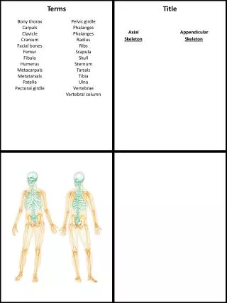
Bony Thorax • Sternum • 12 Ribs • 12 Thoracic Vertebrae • Function • Supports walls of pleural cavity & diaphragm • Volume of cavity able to change during respiration • Protects heart and lungs
Sternum • Flat bone • 6 in in length • Supports clavicles and provides attachment to 1st seven costal cartilages of ribs T2-T3 Sternal Angle T-10 Provides bony landmark for superior liver and inferior heart
12 Rib Pairs • True Ribs • 1-7 • Attached to the Sternum • False Ribs • 8-12 • Do not attach directly to the sternum; attach to costal cartilage of 7th rib • Floating Ribs • 11 and 12 • Attached only to the vertebrae • Number Variation • Cervical Ribs • Articulate with C7 but rarely attach to sternum • Lumbar Ribs • Less Common
Ribs Angle • Oblique plane slanting anteriorly and inferiorly • Anterior ends lies 3-5 inches below the level of the vertebral end. • Angle increases from the rib 1-9 then decreases 9-12.
Ribs • Vary in breadth and length • Facet on head articulates with vertebrae Vertebral End Costal Groove Sternal End Costal arteries, veins, and nerves
Erythropoiesis • Production of red blood cells. • Early Fetus • Mesodermal cells of yolk sac • 3-4 Months to Adolescence • Spleen, Liver, and Skeletal involvement • Adulthood • Vertebrae, Sternum, Pelvis, and Ribs Principal means of delivering oxygen to the body
Bony Thorax Articulations • 8 Joints • Sternoclavicular • Costovertebral (1-12) • Costotransverse (1-10) • Costochondral (1-10) • Sternocostal (1-7) • Interchondral (6-10) • Manubriosternal • Xiphisternal
Sternoclavicular • Only points of articulation between the upper limbs and the trunk • Gliding Joints • Permit free movement Manubriosternal Joint Xiphisternal Joint
Costovertebral and Costotranverse • Costovertebral • Synovial Gliding • Rib Head closely bound to the demifacets and 2 adjacent vertebral bodies • Costotransverse • Synovial Gliding • Tubercle of rib articulates with transverse process of lower vertebra
Costochondral and Sternocostal • Sternocostal • Cartilaginous Synchondosis • No Movement • Articulation between costal cartilages and true ribs • Costochondral • 1st Rib: Cartilaginous Synchondosis • No Movement • 2-7: Synovial Gliding • Freely moveable • Articulation between rib costal cartilages and sternum Sternocostal
Interchondral • Between 6-9 Ribs • Synovial Gliding • Freely moveable • Between 9-10 Ribs • Fibrous Syndesmosis • Slightly moveable
Manubriosternal &Xiphersternal • Cartilaginous Synchondrosis • Little Movement Manubriosternal Joint Xiphisternal Joint
Respiratory Movement • Quiet Respiration • Olique rib orientation changes little • Deep Inspiration • Degree of obliquity decreases • Ribs carried anteriorly, superiorly, and laterally while necks are rotated inferiorly • Deep Expiration • Degree of obliquity increases • Ribs carried inferiorly, posteriorly, and medially while the necks are rotated superiorly
Diaphram • Ribs above diaphram best imaged through air filled lungs • Ribs below diaphram best imaged through upper abdomen • WHY?
Diaphram • Location Changes with Body Position • Upright • Lowest • Supine • Highest • Anterior ends of ribs less sharply visualized in supine position • Repiratory Movement • 1 ½ inches between deep inspiration and deep expiration • Less in hypersthenic • More in hyposthenic
Oblique Projection of Sternum • Why must you do an oblique projection of the sternum versus an AP or PA projection? • Degree of angulation depends on the depth of the chest • Deep Chest • Less angulation • Shallow Chest • More angulation
Which Oblique Position??? • RAO or LAO? • Why?
What technique? • Why?
PA Oblique Projection (RAO)Sternum • Estimate body rotation by placing one hand on patient’s sternum and the other hand on the thoracic vertebrae to act as a guide • Top of IR 1.5 inches above jugular notch Average body rotation is 15-20 degrees
PA Oblique Projection (RAO, LPO)Sternum • Minimal rotation • Sternum projected free from superimposition of the spine • Sternum projected over the heart When would you use an LPO Position?
Lateral Projection (Upright)Sternum • Rotate patients hands posteriorly • Lock hands behind back • Film 24 x 30 cm lengthwise • IR 1.5 inches above jugular notch • Suspend deep inspiration
Lateral Projection (Supine)Sternum • Bring hands above head • Film 24 x 30 cm lengthwise • IR 1.5 inches above jugular notch • Suspend deep inspiration
Pectus Excavatum • Sunken or “caved in” chest • Most common congenital chest wall abnormality in children. • Severity ranges from a moderate indentation to constriction of the internal organs. • Sunken chest appears to be a problem with the sternum or ribs, but the problem is with the cartilage piece that connects each rib to the sternum. This costal cartilage connector is deformed, pushing the breastbone inward.
PA ProjectionSternoclavicular Articulations • IR @ T3 (just posterior to jugular notch) • Arms rest by side of patient with palms up • Turn head toward affected side • Rotates spine slightly away from side being examined • Better visualization of lateral manubrium • Suspend at end of expiration
Sternoclavicular Articulations Bilateral Unilateral No Rotation Slight Rotation
PA Oblique Projection (RAO, LAO)SC Joints • Rotate patient 10-15 degrees • CR perpendicular to SC Joint closest to the IR (T2-T3) S LAO: Left side of interest RAO: Right side of interest L R 15
Ribs • Localize Point of Interest • Anterior Ribs • PA Projection • Posterior Ribs • AP Projection • Axillary Portion of Ribs • Best demonstrated in oblique projection • lateral projection results in superimposition of both sides
Upper Anterior RibsPA Projection • Do you use the same technique as you would for a chest x-ray?
Posterior Ribs: AP Projection • Ribs above diaphram • 1.5 inches above shoulders • Full Inspiration • Ribs below diaphram • Lower edge of IR at iliac crest • Full Expiration
Axillary RibsAP Oblique Projection (RPO, LPO) • 45 degree Oblique • Place affected side closest to the IR • Center affected side midway between midsagittal plane and lateral surface • Abduct arm of affected side and elevate to carry scapula away from rib cage
Axillary RibsAP Oblique Projection (RPO, LPO) • 2 x distance between vertebral column and lateral border affected side visualized • Axillary ribs free of superimposition
Axillary RibsPA Oblique Projection (RAO, LAO) 45 degree oblique • 45 degree oblique • Which is the side of interest? • Why?