Download
1 / 55
560 likes | 888 Vues
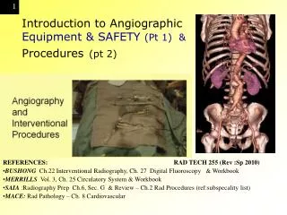
Introduction to Angiographic Equipment & SAFETY (Pt 1) & Procedures (pt 2). REFERENCES: RAD TECH 255 (Rev :Sp 2010) BUSHONG Ch.22 Interventional Radiography, Ch. 27 Digital Fluoroscopy & Workbook MERRILLS Vol. 3, Ch. 25 Circulatory System & Workbook

E N D
Introduction to Angiographic Equipment & SAFETY (Pt 1) & Procedures(pt 2) REFERENCES:RAD TECH 255 (Rev :Sp 2010) BUSHONG Ch.22 Interventional Radiography, Ch. 27 Digital Fluoroscopy & Workbook MERRILLS Vol. 3, Ch. 25 Circulatory System & Workbook SAIA :Radiography Prep Ch.6, Sec. G & Review – Ch.2 Rad Procedures (ref:subspecality list) MACE: Rad Pathology – Ch. 8 Cardiovascular
ANGIOGRAPHY A general term to describe the radiologic examination of vascular structures within the body after the introduction of an iodinated contrast medium.
Special Procedures –Sterile Environment (like OR) The Angio TEAM • Radiologist (Interventional) • CIT/ CV (R. T.) “Angio Tech” • Sometimes more than one • Nurse (Radiology) • Anesthesiologist (if needed) • Other specialists (if needed)
Some history…. • The first angiogram was performed only months after Roentgen's discovery • Which was when? • Two physicians injected chalk or mercury salts into an amputated hand • and created an image of the arteries • Post-mortem injection of mercury compounds, January 1896
Other Technologies/Modailitieswhich demonstrate the vasculature to a greater or less degree • CT • MRI (MRA) • Ultrasound (particularly Doppler) • Nuclear Medicine • are all used to image vessels and each has its advantages and disadvantages • Vessel imaging is a constantly evolving area.
Angiography Equipment:Now primarily uses: DSA: Digital subtraction angiography • This is still considered the gold standard of vessel imaging when other modalities are inconclusive • Now common practice to be considered as an area needing advanced training for: • Radiologist: Interventional • R. T. (CIT, CV) etc ANGIO tech
Technical innovations • image intensification • three-phase generators • rapid film changers • automatic pressure injectors • advanced catheter technology • all helped to establish angiography as an essential diagnostic tool by the 1960s
development of interventional techniques An important offshoot of angiographic imaging • have created a therapeutic technology • Embolization • intra-arterial drug therapy • transluminal angioplasty • are among the procedures that have radically changed and broadened the scope of the diagnostic imaging department
Angiographic Equipment& RoomDesign • Vascular studies usually require a room or suite of rooms • specifically designed to accommodate the sophisticated and accessory equipment needed to perform angiography and interventional procedures
The procedure room should be large enough to accommodate all of the equipment as well as radiologic and ancillary staff • Special procedures sometimes require a general anesthetic that necessitates extra equipment and staff. • These procedures are also more hazardous to the patient and each room must be equipped to deal with emergencies that may occur
Remote Control Center • Remote computerized equipment should also be housed adjacent to the special room. • Although there must be adequate protection for all operators and staff, there must at all times be clear access and view of the patient being examined
EQUIPMENT Needed for Angio* • Biplane C-arm digital imaging • Autoinjector • --syringes, a heating device, • a high-pressure mechanism • a control panel • Image Intensifying screen • Sliding table Rapid film changer (NOW DIGITAL*) • Cut film 6 &Cassette changer /magazine
EQUIPMENT • Multiple Monitors • Remote Contol • Additional Shields • COST: $$$$$$$$$$$$$$ • Angiography equipment • & MRI scanners • are major investments • usually costing between • $750,000 and $1 million DOLLARS
ANGIOGRAPHY EQUIPMENT 1. Puncture Needle Stylet and Cannula large cannula size (1.6mm) 2. Guide Wire --Soft flexible wire with the strength topass through curved vessels (.6 – 1.0)
May have 2 C-arm this Is called what? ________ Should be able to rotate Around the patient
Biplane angiography List 3 advantanges of having 2 tubes simultaneously:
are horizontal only • but with moving or floating capabilities • It is important that during a procedure, a patient can be moved without actually being repositioned, particularly with the catheter in situ. Angiographic table Remote Control – Floating table top to move During the injection of contrast - the table can move to follow the contrast
Angiographic EquipmentSingle or biplane image intensification • A C-arm or U-arm device is preferable • the equipment can be rotated rather than the patient when visualization of the catheter is critical • simultaneous biplane visualization exposures are needed to reduce the number of injections of contrast required
EQUIPMENT • Must be able to pivot 180 degrees • Intensifiers are 12 and 16 inches (larger) • Needs constant potiental – 1500 mA • FOCAL SPOTS • LARGE: 0.6 & 1.2 • SMALL 0.2 & 0.3
Angiographic Equipment Generator: • This must be a three-phase or high-frequency 12-pulse machine and at least 1000 mA to accommodate the rapid, short, and high exposure values required in angiography
Angiographic Equipment Tube Ratings X-ray tube: • High-speed rotating anode tubes. The object of an angiogram is to produce the highest quality radiographs in the shortest time possible • a 0.3 mm small focal spot will produce the best detail • tube rating can be exceeded because of the rapid succession of exposures needed • usual to have a 0.6-mm focal spot tube
HEAT UNITS • Cerebral Study • 1 image / 3 sec for 5 min • 1 image/20 sec for 10 min • HF Generator used (like which phase?) • What is the formula? • What are the heat units for this one study?
automatic film changer • Because of the high pressure of arterial blood flow, the CONTRAST will dissipate through the patient's system quickly, so images must be taken in rapid succession. • An automatic film changer is used because the manual changing of x-ray film can eat up valuable time • Now DSA done digitally
Film changers –NOW OBSOLETE • the ability to move film in rapid succession, • allowing for a number of exposures to be registered each on its own film • There are a number of makes, the most common at present being the Puck system (Siemens) which uses cut film. PUCK-U • Programmable - allowing the operator to vary the speed and the number of films passing through the changer • Speeds vary from 3 to 12 films per second, • These changers can also be single or biplane, allowing for simultaneous exposures.
Film Changer Features (5) Supply magazine. This is a light-tight box that can be filled with film in the darkroom and then attached to the film changer. Transport mechanism. This consists of a series of compression roller devices that moves the film from the supply magazine to a pair of intensifying screens and then to the receiving cassette.
Film Changer Features (5) Compression table. This contains a pair of screens. As soon as the film is positioned between them, they compress the film and the exposure is automatically triggered. As soon as the exposure is complete, the compression is released and the film advanced to the receiving cassette. Receiving cassette. This is the magazine that holds the exposed film. When the examination is complete, the cassette is removed from the changer and taken to the darkroom to be unloaded. It is returned empty to the changer.
Film Changer Features (5) Program selector: • This allows the operator to set speeds and film quantity • to suit the exam being undertaken. Programs can be designed • To fit standard requirements of various procedures.
Film Changer Features (5) • Supply magazine • Transport mechanism • Compression table • Receiving cassette • Program selector
Equipment • Cine radiography. • Fluoroscopy unit with TV monitor: • Single or biplane fluoroscopy units are available. • Video equipment – DIGITAL RECORDING • Other image recording devices: Images can be acquired and stored in a digital format (postprocessing). This is the fundamental principle of DSA.
Pressure Injectors • In most angiographic studies • contrast must be administered at a consistent speed • either faster • as in abdominal angiography • or slower as in lymphangiography SAFETY MEASURES: P 638 LIGHT / ALARM /
Pressure Injectors • Warms contrast – improve viscosity • Piston – motor driven plunger • SAFETY MEASURES (p.638) • Flashing light – audible tone – message • OPERATIONAL ERROR – OMISSION • PREVENTS EXESSIVE PRESSURE FLOW RATE CONTROLLED
Pressure injectorsare motor driven and have the following major components • Control panel where parameters for injections are set. • Motor drive mechanism is the electromechanical device that drives the plunger into the syringe at a specific pressure • Syringes are always removable for sterilization purposes or are disposable. • Heating system maintains the contrast at near body temperature to reduce shock and lower the viscosity of certain contrast media.