Comprehensive Guide to Fracture Diagnosis and Treatment
320 likes | 395 Vues
Learn about fracture diagnosis through clinical features and imaging techniques such as X-rays. Understand the healing process and complications.

Comprehensive Guide to Fracture Diagnosis and Treatment
E N D
Presentation Transcript
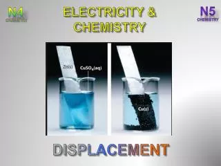
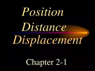
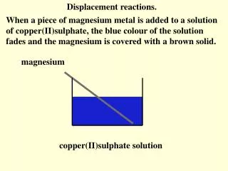
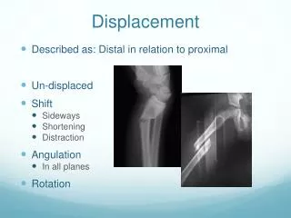
Displacement • Described as: Distal in relation to proximal • Un-displaced • Shift • Sideways • Shortening • Distraction • Angulation • In all planes • Rotation
Fracture Diagnosis • Clinical features • Imaging: Radiology (x-Ray)
Clinical Features • History of Trauma • Symptoms and signs: • Pain • Swelling • Deformity • Bony tenderness • Abnormal movement • Crepitus • Loss of function
Approach - history • Details of injury • Mechanism, force, bleeding, consciousness, … • Details of facture • Deformity, pain, loss of function, .. • Other medical problems • Anti-tetanus status if open injuries • Careful: • Fractures are not always at the site of impact • Some fractures do not need severe force
Approach – clinical exam • General medical condition • should be evaluated to exclude • shock • brain injury • other problems • Vital signs • should be observed and followed up
Approach – clinical exam • Look: • Adequate exposure • General on patient • Local: • Swelling, deformity, bruises, color, … • Special attention is to be paid to wounds
Approach – clinical exam • Feel: • Tenderness, distal pulses, temperature and crepitus on movement • Sensory and motor deficits • Pulse distal to injury • Compartment syndrome • Move: • With care • make sure not to cause more pain or injury • Crepitus &abnormal movement indicates a fracture • Joints distal to the affected area
Approach – clinical exam • Examination of the viscera • Liver and spleen in rib fractures • Urinary bladder and urethra in pelvic fractures • Neurological examination in head and spinal injury
Investigations - Imaging • X-rays: • Low of 2s • Two views: AP and Lateral • Two joints: Above and Below • Two sides: Right and Left • Two occasions • Two Doctors ! • Special views: • Obliques, Tunnel view, skyline, functional flexion / extension • Arthrography: • Shows intra-articular structures • Functional in hip
Imaging • Plain x-ray:(law of twos) • Two views:AP and Lateral AP Lat AP Lat Apley’s System of Orthopedics & Fractures
Imaging • Plain x-ray:(law of twos) • Two views: AP and Lateral • Two joints: joint above and joint below Apley’s System of Orthopedics & Fractures
Imaging • Plain x-ray:(law of twos) • Two views: AP and Lateral • Two joints: joint above and joint below • Two limbs: for comparison • more in children to compare epiphysis Apley’s System of Orthopedics & Fractures
Imaging • Plain x-ray:(law of twos) • Two views: AP and Lateral • Two joints: joint above and joint below • Two limbs: for comparison • more in children to compare epiphysis • Two occasions • e.g. stress fractures • e.g. scaphoid fracture Apley’s System of Orthopedics & Fractures
Imaging • Plain x-ray:(law of twos) • Two views: AP and Lateral • Two joints: joint above and joint below • Two limbs: for comparison • more in children to compare epiphysis • Two occasions • e.g. stress fractures • e.g. scaphoid fracture • Two injuries • e.g. patellar fracture and hip injury • e.g. calcaneal fractures & spine injuries www.jumpintheair.com
Imaging • Plain x-ray:(law of twos) • Two views: AP and Lateral • Two joints: joint above and joint below • Two limbs: for comparison • more in children to compare epiphysis • Two occasions • e.g. stress fractures • e.g. scaphoid fracture • Two injuries • e.g. calcaneal fractures & spine injuries • .....and two Doctors! www.123rf.com/
Imaging • Plain x-ray: (law of twos) • Special views: • Calcaneal view • Shoulder dislocation: axial view • Scaphoid views • Acetabular fractures: 45o tilt views http://osuemed.wordpress.com/
Imaging • CT Scan: • In complex and ntra-articular fractures • In spine • In pelvic and acetabular fractures • In calcaneal fractures www.learningradiology.com
Imaging • MRI • Show associated injuries in spinal fractures • Associated soft tissue injuries – e.g. knee • Hidden fractures: • Subtrochanteric (ST) disruption • Stress (fatigue) fractures • Scaphoid fracture • Suspected avascular necrosis www.highperformancesports.blogspot.com www.bjj.boneandjoint.org.uk
Fracture healing • A broken bone heels because …..it is broken ! Alan Apley
Natural bone healing • Movement at the fracture site initiates a healing process—callus formation • Vascular and cellular response leads to tissue differentiation and mineralization resulting in restoration of mechanical integrity
Natural bone healing http://classes.midlandstech.edu/
Cascade of tissue differentiation Following a Fracture: • Hematoma • Granulation tissue • Connectivetissue • Fibrocartilage • Mineral deposition • Bone
bony bridging Fracture healing • Inflammation • Hematoma • Mesenchymal cells • Soft callus • Granualation tissue • Hard callus • Intramembranous bone formation • Enchondral ossification • Remodeling
Cellular and Vascular Reaction cells haematoma granulation tissue
Tissue Differentiation connective tissue granulation tissue Giemsa
Tissue Differentiation Cascade Masson-Goldner Cartilage formation Mineral deposition Bone
Fracture Healing • Conditions necessary for bone healing: • Good blood supply • Controlled motion • No infection
Fracture Healing • Unfavorable factors • Impairment of blood supply • Infection • Excessive movement • Presence of tumor • Interposition of soft tissue • Any form of Nicotine (smoking) • Bad nutrition
Average healing time • Children: • Upper limb: 3-4 weeks • Lower limb: 2X upper limb (6-8 weeks) • Adults: • Upper limb: 2X children (6-8 weeks) • Lower limb: 2X upper limb (12-16 weeks)
Fracture Treatment • Aim of fracture treatment • aid healing, • in normal position, • avoiding complications
Fracture treatment • Treat the patient, not only the fracture • Reduce the fracture • Immobilize the fracture • Prevents displacement • Alleviates pain • Promotes soft tissue healing • Mobilize the patient • Avoid complications