Download
1 / 33
330 likes | 364 Vues
Learn about the basic components and structure of skeletal muscles, muscle fibers, and how muscles work during exercise. Understand the differences in fiber types and the process of muscle contraction. Discover diagnostic methods and characteristics of muscle fibers.

E N D
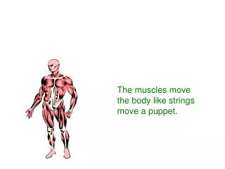
MUSCLES AND HOW THEY MOVE
Learning Objectives wlearn the bacis components of skeletal muscle, muscle fiber wdiscover how muscle functions during exercise wconsider the differences in fibre types wdiagnostika svalových vláken
Key Points Muscle Fiber wAn individual muscle cell is called a muscle fiber. wA muscle fiber is enclosed by a plasma membrane called the sarcolemma. wThe cytoplasm of muscle fiber is called the sarcoplasm. wWithin the sarcoplasm, the T tubules allow transport of substances throught the muscle fiber. wThe sarcoplasmic reticulum stores calcium.
Key Points Myofibrils w Myofibrils are the contractile elemets of skeletal muscle, with several hundred to several thousand composing a single muscle. wMyofibrils are made up of sarcomeres, the smallest functional units of a muscle. wA sarkomere is composed of filaments of two proteins, myosin and actin, which are responsible for muscle contraction. w Myosin is a thick filament with a globular head at one end. wAn actin filament – composed of actin, tropomyosin, and troponin – is attached to a Z disk.
5. The Ca2+ binds to troponin on the actin filament, and the troponin pulls tropomyosin off the active sites, allowing myosin heads to attach to the actin filament. (continued) Excitation/Contraction Coupling 1. A motor neuron, with signals from the brain or spinal cord, releases the neurotransmitter acetylcholine (Ach) at the neuromuscular junction. 2. ACh crosses the junction and binds to receptors on the sarcolemma. 3. This initiates an action potential, providing sufficient ACh. 4. The action potential travels along the sarcolemma and through the T tubules to the SR releasing Ca2+.
Excitation/Contraction Coupling 6. Once a strong binding state is extablished with actin, the myosin head tilts, pulling the actin filament (power stroke). 7. The myosin head binds to ATP, and ATPase found on the head splits ATP into ADP and Pi, releasing energy. 8. Muscle action ends when calcium is actively pumped out of the sarcoplasm back into the sarcoplasmic reticulum for storage.
w Ca2+ ions bind with troponin, which lifts the tropomyosin molecules off the active sites on the actin filament. These open sites allow the myosin heads to bind to them. Key Points Muscle Fiber Action wMuscle action is initiated by a nerve impulse. wThe nerve release Ach, which allows sodium to enter and depolarized, an action potential occurs which releases stored Ca2+ ions.
Key Points Muscle Fiber Action wOnce myosin binds with actin, the myosin head tills and pulls the actin filament so they slide across each other. wMuscle action ends when calcium is pumped out of the sarcoplasm to the sarcoplasmic reticulum for storage. wEnergy for muscle action is provided when thy myosin head binds to the ATP. ATPase on the myosin head splits the ATP into a usable energy source.
Slow-Twitch (ST) Muscle Fibers wHigh aerobic (oxidative) capacity and fatigue resistance wLow anaerobic (glycolytic) capacity and motor unit strength wSlow contractile speed (110ms to reach peak tension) and myosin ATPase w 10–180 fibers per motor neuron
Fast-Twitch (FTa) Muscle Fibers wModerate aerobic (oxidative) capacity and fatigue resistance wHigh anaerobic (glycolytic) capacity and motor unit strength wFast contractile speed (50 ms to reach peak tension) w 300–800 fibers per motor neuron
Fast-Twitch (FTb/FTx) Muscle Fibers wLow aerobic (oxidative) capacity and fatigue resistance wHigh anaerobic (glycolytic) capacity and motor unit strength wFast contractile speed (50 ms to reach peak tension) w 300–800 fibers per motor neuron
Characteristic of muscle fibers Slow-Twitch (ST) SO Fast-twitch (FTa) FOG Fast-Twitch (FTx) FG
SO FG/FOG SPRINTERS AVERAGE CYCLISTS DISTANCE RUNNERS MARATHON RUNERS
DIAGNOSTIC OF MUSCLE FIBERS ► muscle biopsy ► magnetic resonance imaging ► 1MR (one-repetition maximum) and subsequent exrecise with 80%. (1RM) is a functional test of the maximum weight that can be lifted just one time=100% < 8 rep. predominance FG/FOG 8-12 rep. 50%:50% > 12 rep. predominance SO ► Bocso test (jump test)
Muscle Biopsy w Hollow needle is inserted into muscle to take a sample. w Sample is mounted, frozen, thinly sliced, and examined under a microscope. w Allows study of muscle fibers and the effects of acute exercise and exercise training on fiber composition.
w FT fibers have a more highly developed sarcoplasmic reticulum enhancing calcium delivery. (continued) Key Points Slow- and Fast-Twitch Muscle Fibers w Skeletal muscles contain both ST and FT fibers. w ATPase in FT fibers acts faster providing energy for muscle action more quickly than ATPase in ST fibers.
Key Points Slow- and Fast-Twitch Muscle Fibers w Motor units supplying FT fibers are larger (e.g., more fibers per motor neuron) than those supplying ST fibers; thus, FT motor units can recruit more fibers. w ST fibers have high aerobic endurance and are suited to low-intensity endurance activities. w FT fibers are better for anaerobic or explosive activities.
Functional Classification of Muscles Agonists – prime movers, responsible for the movement Antagonists – oppose the agonists to prevent overstretching of them Synergists – assist the agonists and sometimes fine-tune the direction of the movement
Factors Influencing Force Generation wNumber of motor units activated wType of muscle fibers (FT or ST) wMuscle size wInitial muscle length wJoint angle wSpeed of muscle action (shortening or lengthening)
DYNAMOMETRY – muscle strenght testing wDynamometry menas testing of muscles strenght. wStrenght is defined as a peak force of torque development during a maximum voluntary contraction under a given set of conditions. wFor the International System of Units (SI) units for force and torque are the Newton (N) and the Newton meter (N.m)
ISOMETRIC DYNAMOMETRY wIsometric strenght is usually measured as the peak force produced by a maximum voluntary isometric contraction. wDynamometers convert the deformation produced by tension or pressure into srenght (N) wThe dynamometric measurements shoul be made at standardised positions.