Download
1 / 54
540 likes | 695 Vues
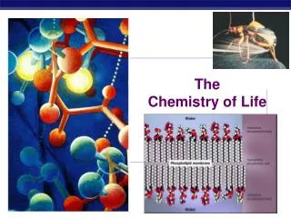
The Chemistry of the cell. M. Saifur Rohman , MD. PhD. FIHA. FICA. Chemical Component of The Cell. Cell Chemistry Is based on Carbon Compounds . A living cell is composed of C, H, N, and O make up nearly 99% of its weight.

E N D
The Chemistry of the cell M. SaifurRohman, MD. PhD. FIHA. FICA
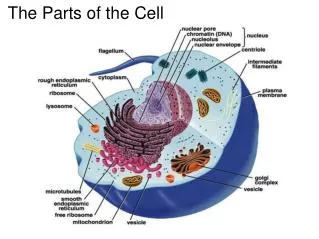
Chemical Component of The Cell • Cell Chemistry Is based on Carbon Compounds . • A living cell is composed of C, H, N, and O make up nearly 99% of its weight. • The most abundant substance of the living cell is water. (70% of a cell's weight), and most intracellular reactions occur in an aqueous environment. • All organisms have been designed around the special properties of water, such as its polar character, its ability to form hydrogen bonds, and its high surface tension.
The Carbon • If we disregard water, nearly all of the molecules in a cell are carbon compounds • The carbon atom, because of its small size and four outer-shell electrons, can form four strong covalent bonds with other atoms. • Most important, it can join to other carbon atoms to form chains and rings and thereby generate large and complex molecules with no obvious upper limit to their size. • The other abundant atoms in the cell (H, N, and O) are also small and able to make very strong covalent bonds
Covalent bond Covalent bonds - form when atoms share electron pairs. • strongest type of bond • tend to form when atoms have 3, 4 or 5 valence electrons • can be nonpolar or polar
Cells Use Four Basic Types of Small Molecules • Certain simple combinations of atoms - such as the methyl (-CH3), hydroxyl (-OH), carboxyl (-COOH), and amino (-NH2) groups - recur repeatedly in biological molecules. • The small organic molecules of the cell have molecular weights in the range 100 to 1000 and contain up to 30 or so carbon atoms. • They are usually found free in solution, where some of them form a pool of intermediates from which large polymers, called macromolecules, are made. • They are also essential intermediates in the chemical reactions that transform energy derived from food into usable forms
Sugars, Fatty Acid, Amino Acid and Nucleotide • All biological molecules are synthesized from and broken down to the same simple compounds. • Biochemical Energetics for cellular reaction and function • Broadly speaking, cells contain just four major families of small organic molecules: the simple sugars, the fatty acids, the amino acids, and the nucleotides. • Although some cellular compounds do not fit into these categories, the four families, and especially the macromolecules made from them, account for a surprisingly large fraction of the mass of every cell
Suggested reading topics • Lodish et al. Chemical foundations in 4th edition Molecular Cell Biology. WH Freeman and Company 2000. Pp 14-49. • Cooper GM. The chemistry of the cell in The cell: A molecular Approach. ASM PRESS. 1997. pp. 39-85.
Amino Acid M. SaifurRohman, MD. PhD. FIHA
Amino Acid • Structure • Classification • Peptide Bond • Force
Amino Acid • Amino acids are molecules containing an amine group, a carboxylic acid group and a side chain that varies between different amino acids. • These molecules contain the key elements of carbon, hydrogen, oxygen, and nitrogen. • Particularly important in biochemistry, where this term usually refers to alpha-amino acids with the general formula H2NCHRCOOH, where R is an organic substituent.
Amino Acids amino group alpha carbon • The general formula for an amino acid • R is commonly one of 20 different side chains • At pH 7 both the amino and carboxyl groups are ionized carboxyl group side chain group
Families of Amino Acids • The common amino acids are grouped according to whether their side chains are: • acidicD, E • basic K, R, H • uncharged polar N, Q, S, T, Y • nonpolar G, A, V, L, I, P, F, M, W, C • Hydrophilic amino acids (uncharged polar) are usually on the outside of a protein whereas nonpolar residues cluster on the inside of protein • Basic or acidic amino acids are very polar and are generally found on the outside of protein molecules
Protein AA : Polymerization • As both the amine and carboxylic acid groups of amino acids can react to form amide bonds, one amino acid molecule can react with another and become joined through an amide linkage. • This polymerization of amino acids is what creates proteins. This condensation reaction yields the newly formed peptide bond and a molecule of water.
Peptide Bonds • Amino acids are joined together by an amide linkage called a peptide bond. • The two bonds on either side of the rigid planar peptide unit exhibit a high degree rotation peptide bonds rotation occurs here
Peptide bond • Protein is a chain of amino acids linked by peptide bonds • Peptide bond • Type of covalent bond • Links amino group of one amino acid with carboxyl group of next • Forms through condensation reaction
Protein Sequence Features • Proteins exhibit far more sequence and chemical complexity than DNA or RNA • Properties and structure are defined by the sequence and side chains of their constituent amino acids • The “engines” of life • >95% of all drugs target proteins • Favorite topic of post-genomic era
Protein Sequence Databases • Where does protein sequence information reside? • Entrez Cross Database Search • http://www.ncbi.nlm.nih.gov/gquery/gquery.fcgi • Swissprot & TrEMBL • http://ca.expasy.org/sprot/ • PIR • http://pir.georgetown.edu/ • As of December 2003, all of this information is integrated into unified protein database called Uniprot. • Uniprot • http://www.pir.uniprot.org/
Protein Structure & Function • Protein structure - primarily determined by sequence • Protein function - primarily determined by structure • Globular proteins: compact hydrophobic core & hydrophilic surface • Membrane proteins: special hydrophobic surfaces • Folded proteins are only marginally stable • Some proteins do not assume a stable "fold" until they bind to something = Intrinsically disordered • Predicting protein structure and function can be very hard --& fun! D Dobbs ISU - BCB 444/544X: Protein Structure & Function
4 Basic Levels of Protein Structure D Dobbs ISU - BCB 444/544X: Protein Structure & Function
primary structure ACDEFGHIKLMNPQRSTVWY Basics of Protein Structure • Primary • Secondary • Tertiary
Primary structure • Sequence of amino acids • Unique for each protein • Two linked amino acids = dipeptide • Three or more = polypeptide • Backbone of polypeptide has N atoms: -N-C-C-N-C-C-N-C-C-N-
Primary Structure & Protein Shape • Primary structure influences shape in two main ways: • Allows hydrogen bonds to form between different amino acids along length of chain • Puts R groups in positions that allow them to interact
Secondary Structure • Local spatial arrangement of amino acids • Description of short-range non-covalent interactions • Hydrogen bonds form between different parts of polypeptide chain • These bonds give rise to coiled or extended pattern • Periodic structural patterns: -helix, b-sheet D Dobbs ISU - BCB 444/544X: Protein Structure & Function
Common Secondary Structure Elements • The Alpha Helix
The a-helix is a common secondary structure element acidic • A helical wheel is a representation of the 3D structure of the a-helix. • Projection of aa side chains onto a plane perpendicular to axis of helix • Hydrophobic arcs stabilize helical interactions • Amphipathic helices are common nonpolar
Common Secondary Structure Elements • The Beta Sheet
Secondary Structure & Protein Folding • Understanding the forces of hydrophobicity: Hydrogen bonds can form with polar side chains on outside of the protein nonpolar side chains polar side chains hydrophobic core contains nonpolar side chains unfolded or partially folded polypeptide folded conformation
Tertiary & Quaternary Structure • Tertiary • Overall 3-D "fold" of a single polypeptide chain • Spatial arrangement of 2’ structural elements; packing of these into compact "domains" • Description of long-range non-covalent interactions (plus disulfide bonds) • Quaternary • In proteins with > 1 polypeptide chain, spatial arrangement of subunits D Dobbs ISU - BCB 444/544X: Protein Structure & Function
Lactate Dehydrogenase: Mixed a / b Immunoglobulin Fold: b Hemoglobin B Chain: a Tertiary Structure
Tertiary Structure Folding as a result of interactions between R groups heme group coiled and twisted polypeptide chain of one globin molecule
Quaternary Structure Some proteins are made up of more than one polypeptide chain Hemoglobin
Force important for protein structure • Ionic bonds • Covalent bonds • Van der waals forces • Hydrogen bonds
Folding of a polypeptide chain Non-covalent amino acid interactions • Hydrogen bonds C=O …. HN • C=O : Glu, Asp, Gln, Asn • NH : Lys, Arg, Gln, Asn, His • OH : Ser, Thr, Glu, Asp, Tyr
Folding of a polypeptide chain Non-covalent amino acid interactions • Ionic bonds COO-….+H3N • COO- : Glu, Asp pKa < 5 • NH3 + : Lys, Arg pKa > 10
Folding of a polypeptide chain Non-covalent amino acid interactions • Van der Waals forces • electrostatic in nature, short ranges • dipole-dipole, ion-dipole etc.
Folding of a polypeptide chain Levels of protein folding • Primary structure (unfolded state) • Secondary structure (-helix, -sheet) • Tertiary structure (domains, subunit) • Quaternary structure (several pp chains) • Intra- and intermolecular disulfide bonds
Suggested reading topics • Lodish et al. Protein structure and function in 4th edition Molecular Cell Biology. WH Freeman and Company 2000. Pp 50-99.
Protein Synthesis, Processing and Regulation M. SaifurRohman, MD. PhD. FIHA
Protein Synthesis Intracellular • In cells, this reaction does not occur directly; instead the amino acid is first activated by attachment to a transfer RNA molecule through an ester bond. • This aminoacyl-tRNA is then a substrate for the ribosome which catalyzes the attack of the amino group of the elongating protein chain on the ester bond. • All proteins made by ribosomes are synthesized starting at their N-terminus and moving towards their C-terminus
Protein synthesis • DNA • mRNA (transcription) • Protein (translation)
Post-Translational Modification • New polypeptides usually fold themselves spontaneously into their active conformation. However, some proteins are helped and guided in the folding process by chaperone proteins • Many proteins have sugars, phosphate groups, fatty acids, and other molecules covalently attached to certain amino acids. Most of this is done in the endoplasmic reticulum. • Many proteins are targeted to specific organelles within the cell. Targeting is accomplished through “signal sequences” on the polypeptide. In the case of proteins that go into the endoplasmic reticulum, the signal seqeunce is a group of amino acids at the N terminal of the polypeptide, which are removed from the final protein after translation.
Posttranslational modifications Chemical alterations after protein synthesis • May alter activity, life span or cellular location • Chemical modification • Acetylation, phosphorylation, glycosylation • Processing • Proteolytic (in)activation, selfsplicing
Protein processing and regulation Protein modification : becoming a functional protein • Folding • Proteolysis • Glycosylation • Signal sequence