Download
1 / 46
570 likes | 950 Vues
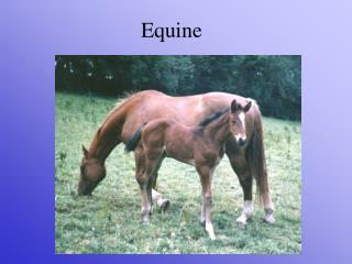
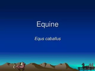
The Equine Foot and Physiologic Trimming. Brendan Kraus, DVM With thanks to Dr. Steve O’Grady. The Equine Foot. The foot has numerous functions Supporting the horses weight Dissipating the energy of impact Protecting the structures within the hoof capsule Providing traction.

E N D
The Equine Foot andPhysiologic Trimming Brendan Kraus, DVM With thanks to Dr. Steve O’Grady
The Equine Foot • The foot has numerous functions • Supporting the horses weight • Dissipating the energy of impact • Protecting the structures within the hoof capsule • Providing traction
The Equine Foot • The equine foot must follow the laws of physics • Trimming and shoeing affects both the external hoof capsule and the internal structures of the foot • Many foot lamenesses can be prevented or or treated with good farrier care
The Equine Foot A “functional” foot is comprised of several key components • A parallel hoof-pastern axis • A thick hoof wall • Adequate sole depth • A solid heel base with heels of the hoof capsule extending to the base of the frog • Equal growth rings from toe to heel
Anatomy of the Foot • Bony Structures • Distal end of Second Phalanx • The Distal Phalanx (coffin bone) • The Navicular Bone
Anatomy of the Foot • Coffin Joint • Formed by middle phalanx, distal phalanx, navicular bone, and deep flexor tendon supporting • Center of Articulation of leg
Anatomy of the Foot • Navicular Bone • Increases the size of the articular surface of the joint • Maintains a constant angle of insertion of the deep flexor tendon on the coffin bone • Navicular bone’s position makes this region susceptible to a wide range of forces and movements
Anatomy of the Foot • Soft Tissue Structures • Digital cushion • Ungual cartilages • Deep flexor tendon • Lamellae • These structures offer anti-concussive function during weight bearing and dissipate impact energy during landing
Soft Tissue Structures • Lamellae
Anatomy of the Foot • Bony Structures • Hoof Complex • Weight bearing epidermal structures • Hoof Wall • Bars • Sole adjacent to sole/wall junction • Soft Tissue anti-concussive structures • Frog • Digital cushion • Ungual cartilages • Deep flexor tendon • Lamellae
Hoof Complex-Weight Bearing Structures • Hoof capsule is thickest at toe for stiffness • It decreases thickness toward the heels • Allows for heel flexibility and expansion • The hoof wall inflicts at an acute angle at heels and extends under the foot to form the bars • The hoof wall, bars, and sole form the heel base • The sole should be concave to allow it to flatten in weight bearing • Sole depth should be 15mm
The Palmar Foot • The structures that comprise and form the palmar foot are often the limiting factor when trying to achieve and maintain good hoof conformation or shape • Frog, digital cushion, ungual cartilages • The conformation of the palmar foot will be contingent on the accumulated mass of these structures • Sufficient mass is not always possible
Palmar Foot Mass • The structures that comprise the palmar foot are influence by • Genetics • Improper Development or Maturity • Continuous Repetitive Overload (immaturity) • Inappropriate farrier practices
Hoof Balance • What is Hoof Balance? • The Ideal Shape or Conformation of the Foot • “Hoof Balance” Lacks an Intrinsic Definition • An Option to the term “Hoof Balance” is to use a set of biomechanical landmarks as guidelines • This set of guidelines can be applied to every horse
“Hoof Balance”Guidelines • The Hoof-pastern axis • The center of articulation of coffin joint • The entire limb rotates around this point • The extension of the heels to the base of the frog
Hoof-Pastern Axis • The first guideline when trimming the foot • When the horse is standing with the cannon bone perpendicular to the ground and viewed from the side, the HPA should form a straight line.
Hoof-Pastern Axis • The line should pass through the middle of the phalanges from the fetlock to the ground.
Hoof-Pastern Axis • The changes to the HPA are associated with two common hoof capsule distortions • Broken-Back HPA • A low or under-run heel • Often has excessive toe length • The low Heel generally has a lack of soft tissue mass so the foot is unable to dissipate energy during landing
Broken-Back HPA • This type of foot conformation is quite common • One study found normal performance horses to be 52% affected • 77% of horses with foot related lameness was found in another study
Broken-Back HPA • A low hoof angle places the joints of the digit in hyperextension, promotes load bearing on the heels, and increases strain on the deep flexor tendon. • This excess load may cause increased stress on the navicular apparatus and the associated soft tissues • If a horse is experiencing pain in the heels, it will land toe first which can lead to bruising. • Long toe/Low Heel may contribute to chronic heel bruising, coffin joint inflammation, quarter and heel cracks, interference, and compromised heel circulation
Hoof-Pastern Axis • The changes to the HPA are associated with two common hoof capsule distortions • Broken-Forward HPA • The heels grow long • The long heels will bypass the soft tissue structures of the palmar foot causing energy of impact to be transferred directly to bone • Promotes load bearing on the toe and dorsal margin of the coffin bone • Severe form classified as “Club foot” which has a very high hoof angle as well as misalignment of the bones
“Hoof Balance”Guidelines • The Hoof-pastern axis • The center of articulation of coffin joint • The entire limb rotates around this point • The extension of the heels to the base of the frog
Center of Articulation • A vertical line drawn down from the center of the coffin joint should bisect the widest part of the sole • This point remains relatively constant regardless of the shape or length of the heels or toe • Since this is the pivot point of the foot, the distance from the line to the heels and toe should be equal
Center of Articulation The trimmed foot is as wide as it is long
“Hoof Balance”Guidelines • The Hoof-pastern axis • The center of articulation of coffin joint • The entire limb rotates around this point • The extension of the heels to the base of the frog
Heels to Base of Frog • The hoof capsule contains both bony and soft tissue structures. • For these structures to be able to work together, they both need to be enclosed within the hoof capsule in the same plane • This necessitates that the hoof wall at the heels extend to the base of the frog
Heels to Base of Frog If the heels or shoe branch stop short, the concussive structures are no longer in the same plane as the sole
Heels to Base of Frog • If the heels are left too long, or are allowed to grow forward, the function of the soft tissue structures are assumed by the bones • If a foot has limited soft tissue mass, the heels cannot be trimmed to the base of the frog • This necessitates that some form of farriery extend to the base of the frog
Use of Radiographs (x-rays) • A diagnostic tool • An aid in assessing internal landmarks • A blueprint of the foot for veterinarians and farriers
Shoes • The shoe should be an extension of the correctly trimmed foot
Summary • Adherence to the basic principles of farriery is essential to maintaining hoof health and continuous soundness • Becoming familiar with these basic landmarks will enable an approach to trimming the equine foot that is standardized and repeatable
Questions? Tonight’s material can be reviewed at equipodiatry.com