Download
1 / 53
530 likes | 1.2k Vues
Cardiovascular System L- 3 Physiology of the Heart and Blood Circulation. Dr Than Kyaw 20 Feb 2012. Goals. Understanding:-. Functional anatomy of the heart , - pericardium , myocardium, cardiac muscle. Excitability and conductive pathway of the heart, heart beat

E N D
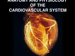
Cardiovascular SystemL- 3 Physiology of the Heart and Blood Circulation Dr Than Kyaw 20 Feb 2012
Goals Understanding:- Functional anatomy of the heart, - pericardium, myocardium, cardiac muscle. Excitability and conductive pathway of the heart, heart beat Explain the functioning of the valves of the heart and how they relate to the heart sounds. Blood flow, blood pressure
Cardiovascular System (CVS) The system consists of: - Fluid (blood) - Vessels (arteries, veins, capillaries) - A pump (heart)
Plasma and its constituents In Lecture 2 – a little about plasma has been discussed Plasma = Blood minus formed elements (remember hematocrit) Serum = plasma minus most of cloting factors (supernatant yellow fluid that remains after a clot forms; contains antbody fractions of the blood)
Plasma and its constituents • Plasma - 92% water; 8% other substances • Osmolality = 290mOsm/kg at normal body temperature • Na+ and Cl – ions contribute the most to total osmolality • Albumin and globulin, 2 major proteins synthesized in liver - many small compounds and electrolytes bind to albumin, circulate in bound forms preventing their rapid loss in urine • Albumin and other large molecules do not readily pass capillary walls; provide an effective osmotic force - prevent excessive fluid loss from capillaries to outside
Plasma and its constituents • - and -globulins - functions as albumins - body defense - as precursor enzymes in blood clotting • -globulin (synthesized in cells of immune system) - immunoglobulins: IgG, IgE, IgA, IgM, IgD - IgG most abundant • In some animals immunoglobulins pass placenta; give immunity to the newborns • Some animals through colostrum to newborns
Location of the Heart • Hollow muscular structure located in the thorax • Large arteries & veins – continuous with the heart at its base • Base directed upward and forward; apex, the opposite end, directed downward and backward • in pericardial space (sac)
Covering of the heart - Pericardium 1. Fibrous pericardium - tough, collagenous 2. Parietal pericardium (lines fibrous pericardium) 3. Visceral pericardium (epicardium) adheres to the heart surface 4. Pericardial space filled with small amount of pericardial fluid important for lubrication of the heart for near-continuous motion
Structure of Heart Wall (myocardium) • Epicardium = visceral Pericardium (serosa) • Myocardium: muscle tissue + C/T + blood vessels + ? • Endocardium: - simple squamous epithelium continuous with endothelia of blood vessels - because of its smooth surface - reduce friction - minimize resistance for blood flow and thus lower energy requirement What will happen when - Endocarditis – inflammation of endothelial lining of the heart Valvularendocarditis – endocarditis involving inflammation of endothelium of the valves
Myocardium • Myocardium - forms the wall + compartments (chambers) • Muscles - arranged in a manner so that when they contract the blood is ejected from the chambers • Right side and left side chambers; each side has an atrium and a ventricle • Each atrium has an extension k/s auricle (to conserve space) • Atria receive blood from the veins and the ventricles receive blood from the atria • Right and left ventricles pump blood from the heart through the pulmonary trunk and aorta, respectively
Left vs. Right Ventricle Muscles Left: high pressure pump (Why?) Right: low pressure pump right chamber is thinner walled than left Ventricles separated by interventricular septum
Cardiac Muscle • Striated, aerobic, interwoven, autorhythmic • Intercalated discs - gap junctions, strong junctional complex (desmosomes)
Properties of the cardiac muscle • Excitability • It is the ability of the cardiac muscle to respond to a stimulus. • The index for excitability is Chronaxie. • the minimum time required for excitation of a nerve or muscle when the stimulus is double the minimum (threshold) necessary to elicit a basic response.
Properties of the cardiac muscle Electrophysiology of the heart: I. Resting membrane potential (RMP) = - 60 MV This is because cardiac muscle is more permeable to Na+ ions Cardiac muscle, being less excitable, has higher Chronaxie than skeletal muscles(RMP of skeletal muscle = - 90 mv and smooth muscle = - 70 mv) II. Prolonged action potential Due to the plateau caused by opening of slow Ca+ channels which prolongs depolarization and thus mechanical shortening of the cardiac muscle occurs.
Properties of the cardiac muscle Contractility - The cardiac muscle has the ability to contract isometrically and isotonically Isometric contraction: The length remains constant but the tension increases e.g. early phase of ventricular systole Isotonic contraction: The tension remains constant while the length shortens e.g. late phase of ventricular contraction Contractility obeys "All or None Law" and "Starling Law" All or None Law: the strength by which a nerve or musclefiber responds to a stimulus is not dependent on the strength of the stimulus. If the stimulus is any strength above threshold, the nerve or muscle fiber will give a complete response or otherwise no response at all.
A. Resting B. Isometric contraction; m/s no change in length, sarcomeres shorten, stretching the series elastic elements C. isotonic contraction; the contractile elements shorten, stretching the series elastic elelments, before they develop tension to lift the load. D. Muscle begins to shorten when contractile elements shorten further.
Starling Law: - The further the stretch of the muscle fibers, the stronger is the contraction. - Up to a certain limit beyond which the muscle fibers can no longer contract stronger even with greater stretch of the muscle fibers. Conductivity -- Ability of the myocardial fibers to spread conduction along the conduction system all over the heart. -- Conductivity myocardial fibers vary but generally have a high conductive velocity.
Metabolism of cardiac muscle Cardiac Muscle • Heart metabolism is different from skeletal muscle in 3 ways. • Heart muscle can function only under aerobic conditions. Heart muscle cells are rich in mitochondria facilitating aerobic respiration. • Heart muscle - unableto store glycogen. • Fatty acids are the preferred fuel of the heart. Glucose is the least favored fuel. • Ketone bodies and lactate are used under stress when the energy demand is high.
Rythmicity -- Ability of the heart to contract with regular intervals of relaxation -- the distance between consecutive beats or myocardial contractions is almost equal or the duration of relaxation is almost the same Autorythmicity -- the ability to conduct and contract regularly without an external stimulus. -- In spite of that, the heart is still being adjusted by ANS and hormones to adjust cardiac muscle function in certain conditions e.g. stress
Autorythmicity - SinoatrialNode (S.A): 60 – 80 charge/min (natural pacemaker of the heart) - AtrioventricularNode (A.V): 40-60 charges/min- BundleofHis: 30 – 40 charges/min- Purkinjéfibers: 15 charges /min (incompatible with life) - If S-A node fails to initiate a stimulus, any of the other areas will initiate the stimulus and will be considered an ectopic pacemaker for the heart.
Heart valves • Atrioventricular (A-V) valves - Right side = tricuspid valve - Left side = bicuspid (mitral) valve - A-V valves prevent expulsion of ventricular blood into the atria when the ventricles contract. • Tendency of A-V valves eversion is prevented by cords (chordaetendineae) attached to the free margin of the cusps at one end and to small muscles (papillary m/s) at the other end that extend from the myocardium • Papillary m/s contraction is synchronized with the myocardial contraction so that tension to the chordaetendineae is approximately timed
Heart valves • Back flow of blood just ejected from the ventricles is prevented by valves located at the exits of the arteries from the ventricles • Pulmonarysemilunar valve - for pulmonary trunk • Aortic semilunar valve – for aorta • All heart valves ensure unidirectional blood flow
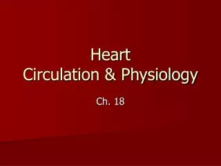
The Mammalian Heart • Showing direction of blood flow
Blood flow through the Heart Systemic and Pulmonary
Blood flow through the Heart • Pulmonary vein • Venous Blood • Blood from tissues in forward part of the body • Cranial • vena cava • R - A • L - A • Caudal • vena cava • R - V • L - V • Blood from tissues in rear part of the body • Aorta • Lung Fig 21.6
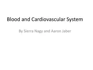
Structure and Function of Valves = Mitral valve 4 sets of valves Prevent backflow of blood Close passively under blood pressure Heart sounds produced by valve closure
Support for AV valves: valves are restrained by chordae tendinae which are in turn attached to papillary muscles (prevention of backflow!) picture taken from R ventricle, looking toward R atrium
Heart Valve Prolapse Mitral Valve Prolapse • Most common cardiac variation (5-10% of population) • Mitral valve cusps do not close properly • Regurgitation during left ventricular systole
Special circulation: coronary , pulmonary & cerebral Leaves by passing pulmonary semilunar valves into pulmonary trunk and to the lungs to be oxygenated Blood from body enters right atrium Passes tricuspid valve into right ventricle Returns from the lung by way of pulmonary veins into the left atrium Leaves left ventricle past aortic semilunar valves into aorta From left atrium past bicuspid valve into left ventricle • Distributed to rest of the body
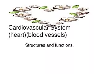
Coronary circulation • circulation of blood in the blood vessels of the heart muscle (the myocardium) • vessels - deliver oxygen-rich blood to the myocardium • coronary arteries • cardiac veins. - Superficial coronary arteries - epicardial coronary arteries • when healthy - capable of autoregulation to maintain coronary blood flow • at levels appropriate to the needs of the heart muscle. • relatively narrow vessels; commonly affected by atherosclerosis ; become blocked; • cause - angina or a heart attack. • - Deep coronary arteries - subendocardial. • Coronary arteries – k/s "end circulation“ • - because the only source of blood supply to the myocardium • - very little redundant blood supply • - blockage of these vessels can be so critical
Coronary Circulation Coronary arteries: first branches off the ascending aorta.
coronary veinscoronary sinusright atrium (inferior to opening of inferior vena cava) posterior view
Coronary heart diseaseCoronary artery disease (CAD)Arteriosclerotic heart disease (CHD) a narrowing of the small blood vessels supply of blood and oxygen to the heart slow down or stop
Coronary Artery Disease (CAD) due to ? consequences ? PET scan (Positron Emission Tomography ) the brighter the color, the greater the blood flow through tissue
Myocardial Infarction (MI) • ~ 1.3 mil MIs / year in US • Most commonly due to severe CAD (coronary thrombosis) • Ischemic tissue degenerates → nonfunctional area = infarct • Predisposing factors?
Conducting System of the Heart Specialized muscle cells (autorhythmic cells) conduct APs to time and synchronize the action of the chambers SAnode– Also k/s pacemaker, located on the wall of right atrium - Initial stimulus - spontaneously depolarizes most rapidly - initiate heart beat - transmits action potential across the right and left atria - contraction of atria emptying the contents (blood) into the ventricles
Conducting System of the Heart AVnode - when the impulse reaches AV node, it travels down along the AVbundle (bundle of His) within the septum Bundle of His branches, one of which supplies each ventricle where they branch into Purkinje fibers Purkinje fibers reflect up external walls of ventricles and stimulate contraction of cardiac muscle cells as a unit. The blood in the ventricles – pumped into pulmonary artery and aorta to circulate to pulmonary and systemic circuits.
Conducting System of the Heart - Atria and ventricles - separatedby a fibrous ring surrounding the AV valves. • It acts as an insulator. • Therefore the impulse that spreads throughout the atria does not spread to the ventricles and vice versa • This permit independent contractions of atria and ventricles • i.e, the ventricles are filled during their relaxation by the contraction and emptying of the atria. • AV bundle fibers - smaller diameter than other Purkinje fibers - conductivity - 10% slower than cardiac muscle fibers. • Ventricular m/s - thicker than atria and conduction distance is greater – therefore to achieve coordinated contraction of muscle fibers of ventricles , it is essential to have a greater velocity of conduction which is provided by Purkinje fibers.
Conducting System of the Heart Cardiac muscle – contract more slowly than skeletal m/s - longer refractory period (the period during repolarization when a stimulus cannot evoke another depolarization. Autorhythmic stimulation may be modified by sympathetic and parasympathetic nerves changing the nature of heart beat in terms of frequency and force of contraction
Cardiac cycle and Heart Beat Cardiac cycle = the sequence of events that occurs during one complete heart beat. 2 phases – systole (contraction) diastole (relaxation) 2 phases are continuous - assigned periods are arbitrary - The two atria are in systole and diastole together as are the two ventricles. After ventricular systole and during ventricular diastole: the following sequence of events occurs
Cardiac cycle • Atria filled by blood from Venaecavae and pulmonary veins; vol. & pressure (occurs during ventricular systole) AV valves open when arterial pressure exceeds the ventricular pressure (occurs at the beginning of ventricular diastole) • Blood flows into relaxed ventricles (abt 70% filled) • Atria contract (filling of ventricles complete) • Atria relax and begin filling. • Ventricles begin contraction; AV valves closed b/c ventricle pressures exceeds atrial pressures • Continued contraction of ventricles • Semilunar valves open • Blood ejected from ventricles. • Ventricles begin to relax. • Atrial pressures begin to exceed the ventricular pressures and semilunar valves closed.
Heart sounds Two heart sounds by auscultation: - 1st sound = Lub = closure of aterioventricular valves when ventricles contract 2nd sound = dub = closure of semilunar valves when ventricles start to fill following contraction (3rd and 4th sound may be detected on ECG; 3rd sound due to flowing of blood into ventricles and 4th sound due to atrial contraction) Murmurs: abnormal heart sounds resulted from valve disorders.
Factors that affect heart rate Physiological - excitement - muscular exercise (activity, work, exercise) - high environmental temperature - digestion - sleep - high altitude - smoking - some food or drinks – e.g. caffeine (coffee, tea) - respiration (slight changes) - metabolic rate