Download
1 / 51
510 likes | 707 Vues
Principles of Skeletal Muscle Adaptation. Brooks ch 19 p 430- 443. Outline. Myoplasticity Protein turnover Proposed regulatory signals for adaptation Fiber Type Training Inactivity. Myoplasticity.

E N D
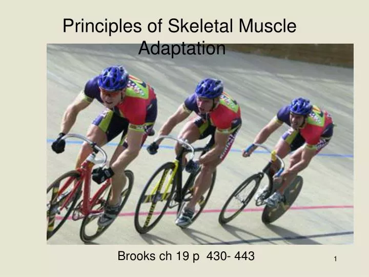
Principles of Skeletal Muscle Adaptation Brooks ch 19 p 430- 443
Outline • Myoplasticity • Protein turnover • Proposed regulatory signals for adaptation • Fiber Type • Training • Inactivity
Myoplasticity • Altered gene expression - results in an increase or decrease in the amount of specific proteins • tremendous potential to alter expression in skeletal muscle • The adaptations result in more effective aerobic or resistance exercise • This is the molecular basis for training adaptations
Myoplasticity • Chemical messengers have an important role in stimulating adaptations to exercise training • Chemical messengers respond to physical and mechanical stress, neural signals, metabolic, bioenergetic, hypoxic and temperature signals resulting from aerobic or resistance exercise • 20% of skeletal muscle is protein,balance is water, ions... • All proteins can be regulated by altering gene expression • Fig 19-2cascade of regulatory events impacting gene expression • Muscle gene expression is affected by changes induced by loading state and the hormonal responses occurring with exercise • Regulation occurs at any level from transcription to post translation • transcription factors interact with their response elements to affect promotion of various genes
Myoplasticity cont. • Fig 19.2 continued • Hormones bind to nuclear receptors (HR) and interact with DNA at Hormone response elements (HRE) to affect transcription • Activity (loading) changes levels of certain Transcription Factors (TF) (c-fos, c-jun, CREB, MAPK) • Activity also changes levels of circulating hormones • myoplasticity - change either quantity (amount) or quality (type) of protein expressed • Eg. Responses to training • Quantity - hypertrophy (enlargement)- increased protein in fiber • Quality - repress gene for fast II b myosin HC, turn on fast IIa myosin HC
Protein turnover • Protein Turnover reflects 1/2 life of protein - time frame for existence • protein transcribed (DNA-mRNA) • translated then degraded • level of cell protein governed by • Balance of synthesis / degradation • precise regulation of content through control of transcription rate • and/or breakdown rate • Mechanism provides the capacity to regulate structural and functional properties of the muscle • applies to proteins involved in; • Structure, contraction, and transport • as well as enzymes involved in metabolism
Adaptation • Sk ms adaptations are characterized by alterations in functional attributes of muscle fibers through; • Morphological, Biochemical and Molecular variables • adaptations are readily reversible when stimulus is diminished or removed (inactivity) • Fig 19-3 - many factors can modify microenvironment of fiberwhich in turnregulates gene pool expression • changes can lead to altered rates of protein synthesis and degradation • changing content or activity of proteins • Microenvironment includes the intracellular milieu and immediate extra-cellular space
Signals for Adaptation • Insufficient energy intake • Leads to protein degradation for fuel • anorexia, sarcopenia • Increased cortisol • inhibits protein synthesis by blocking AA uptake into muscle, blocks GH, IGF-1 and insulin actions • Stimulates protein degredation • nutrition also influence hormones • Insulin - anabolic • power developed by motor unit • Recruitment and load on fibers • specific responses result from; • Reduced power, sustained power, or high power demands • May utilize myogenic regulatory factors to stimulate transcription
Signals for Adaptation • Hormones - independent of nutrition • thyroid hormone - gene expression at all levels pre and post transcriptional and translational • Eg myosin heavy chain, SR Ca++ pump • Importance with training is unclear • IGF-1 - insulin like growth factor 1 • mediates Growth Hormone effects • Stimulates differentiation and incorporation of satellite cells • Muscle release of IGF-1 independent of ciculatory IGF-1 release induced by GH
Signals for Adaptation • GH stimulates liver release of IGF-18-30 hours post exercise • muscle release of IGF-1 induced by RE • more important for muscle specific adaptations • Fig 19-4 • Exerts Autocrine/paracrine effects • MGH - mechanogrowth factor • Training inc IGF-1 mRNA expression • Inc GH dependant /independent release
Signals for Adaptation • Endurance Training • small rise during exercise • Greater rise when training above lactate inflection point • GH – positive correlation between GH and aerobic fitness • GH may be mediator of increased O2 and substrate delivery and lipid utilization by exercising muscle • Improves FFA oxidation - stimulating lipolysis during but mainly after exercise • Reduces glucose uptake after exercise by inhibiting insulin action • GH may also play a role in improved thermoregulation, conversion of muscle fibers to more oxidative and up-regulation of oxidative genes to improve mitochondrial function that occur with endurance training
Signals for Adaptation • Resistance Training (RE) • Testosterone and GH - two primary hormones that may affect adaptations to RE • Both Inc secretion with training • Testosterone - inc GH release • Inc muscle force production - Nervous system influence • Direct role in hypertrophy still being investigated • IGF-1, T and RE required to stimulate satellite cells and result in hypertroyphy and increased strength. • Muscle damage from RE also stimulates satellite cell proliferation.
Metabolic Regulation • Many proposed factors related to fatigue and the intracellular environment • Calcium concentration increases 100 fold with muscle stimulation • Increase is recruitment dependant and motor unit specific - • influence varies with frequency and duration of stimulation and cellular location of calcium • Calcium influences transcription through kinase cascades and transcription factors • stimulating muscle growth in response to high intensity activity (hypertrophy) • Calcium - Calmodulin Dependant protein kinase • Unknown whether calcium plays an essential role in hypertrophy
Metabolic Regulation • Redox state of cell is influenced by activity level. • The content of Reactive oxygen species (ROS) increases with duration of activity (endurance) • ROS along with hypoxia and low cellular engergy activate a cascade of transcription factors stimulating growth of mitochondria • increase aerobic enzyme content (more study required) • May have influence in conjunction with Thyroid hormone on mitochondrial DNA – up-regulating mitochondrial biogenesis and beta oxidation
Acute Exercise and Glucose metabolism • Insulin and muscle contraction stimulate an increase in glucose uptake into muscle • via different intracellular pathways (fig 1) • Glucose Transporters (GLUT 4) migrate to cell surface from intracellular pools • facilitated diffusion of glucose into cell • Type II diabetes may involve errors in insulin signaling or the downstream stimulation of GLUT 4 migration • With exercise, delivery, uptake and metabolism of glucose needs to increase
Acute Exercise and Glucose metabolism • Muscle contraction increases Ca++ and AMPK (AMP-activated protein kinase) • Ca++ may act through CAMK (calmodulin-dependant protein kinase) or calcineurin • Acute Ca++ stimulates migration of GLUT 4 to surface • AMPK - regulated by intracellular ratios of ATP:AMP and CP:creatine • Acute AMPK- stimulates migration of GLUT 4 to surface
Chronic exercise and Glucose metabolism • Chronic increases in Ca++ maystimulate transcription factors • MEF2A, MEF2D, NFAT • Levels of GLUT 4 protein and mitochondrial enzymes observed to increase in laboratory studies • AMPK - regulated by intracellular ratios of ATP:AMP and CP:creatine • Chronic exposure to an AMPK analog (AICAR) results in increased GLUT 4 protein expression, HK activity in all muscle cells • CS, MDH, SDH, and cytochrome c increased in fast twitch muscle only • Endurance training produces similar results to those indicated with Ca++ or AMPK • Increased GLUT 4 protein content • increases capacity for glucose uptake from circulation • may improve glucose tolerance during early stages of the development type 2 diabetes by stimulating insulin sensitivity or increasing GLUT 4 migration
Phenotype • When protein structure of muscle is altered - the phenotype changes • Phenotype is outwardly observable characteristics of muscle • Slightly different versions of proteins can be made - isoforms • This reflects underlying genes (genotype) and their potential regulation by many factors (eg exercise) • altered phenotypes - affect chronic cellular environment and the response to acute environmental changes (training effects) • eg. Receptors, integrating centers, signal translocation factors and effectors are modified in content or activity- • signaling mechanisms are not fully understood - molecular biology is helping elucidate control pathways
Hereditability of Fiber TypesPercent Slow Twitch Fibers Identical Twins Fraternal Twins Twin A Twin A 0 20 40 60 80 0 20 40 60 80 0 20 40 60 80 0 20 40 60 80 Twin B Twin B
Muscle Fiber Types • Elite athletes - specialized fiber typing • sprinters II b, endurance athletes type I • Fig 19-5 - elite - specialized at the ends of the fiber type spectrum • Training studies - alter biochemical and histological properties - but not fiber type distinction • Fiber typing is according to myosin heavy chain isoform • evidence, however, that intermediate transitions can occur in MHC expression • not detected with conventional analysis techniques
Endurance Adaptations • Occurs with large increase in recruitment frequency and modest inc in load • minimal impact on X-sec area • significant metabolic adaptations • Increased mitochondrial proteins • HK inc, LDH (dec in cytosol, inc in mito) • 2 fold inc in ox metabolism • degree of adaptation depends on pre training status, intensity and duration
Endurance Adaptations • Table 19-1 Succinate DH (Krebs) • response varies with fiber type - involvement in training • inc max blood flow, capillary density, and potential for O2 extraction
- Increases in oxidative enzyme mRNA several hours after endurance exercise - no change in cytoskeletal factors (Titin)
Adaptations to Resistance Training • Inc recruitment frequency and load • Hypertrophy - inc X-sec area • Increase maximum force (strength) • Fig 17-31b - Force velocity after tx • move sub max load at higher velocity • enhance power output (time factor)
Adaptations to Resistance Training • Fiber type specific adaptation • inc X-sec area of both type I and II • Fig 19-6 (5-6 month longitudinal study) • Type II - 33% , Type I-27% increase
Adaptations to Resistance Training • Fastest MHC’s repressed • inc in expression of intermediate MHC isoforms - some Type II x shift to II a • mito volume and cap density reduced • Fig 19-7 - 25 % dec in mito protein
Adaptations to Resistance Training Fig 19-8 - cap density dec 13%
Inactivity / detraining • Aging, space flight, bed rest, immobilization from injury • large reduction in recruitment frequency and /or load • Significant reduction in metabolic and exercise capacity in 1-2 weeks • Complete loss of training adaptations in a few months • VO2 max dec 25 % • Strength improvement lost completely • Adaptations • reduction in ms and ms fiber X-sec area - decrease in metabolic proteins • Fig 19-10