STRUCTURE OF SKELETAL MUSCLE
270 likes | 651 Vues
بسم الله الرحمن الرحيم. STRUCTURE OF SKELETAL MUSCLE . Dr.Mohammed Sharique Ahmed Quadri Assistant Professor, Physiology. Objectives. By the end of this lecture, you should be able to:

STRUCTURE OF SKELETAL MUSCLE
E N D
Presentation Transcript
بسم الله الرحمن الرحيم STRUCTURE OF SKELETAL MUSCLE Dr.Mohammed Sharique Ahmed Quadri Assistant Professor, Physiology
Objectives By the end of this lecture, you should be able to: • Draw and label a skeletal muscle at all anatomical levels, from the whole muscle to the molecular components of the sarcomere. At the sarcomere level, include at least two different stages of myofilament overlap. • Diagram the structure of the thick and thin myofilaments and label the constituent proteins. • Describe the functional importance of the subunits.
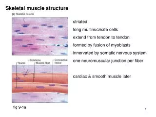
Structure of Skeletal Muscle • Skeletal muscle consists a number of muscle fibers lying parallel to one another and held together by connective tissue • Single skeletal muscle cell is known as a muscle fiber • Multinucleated • Large, elongated, and cylindrically shaped • Fibers usually extend entire length of muscle
Structure of Skeletal Muscle • Myofibrils • Contractile elements of muscle fiber • Regular arrangement of thick and thin filaments • Thick filaments – myosin (protein) • Thin filaments – actin (protein)
Viewed microscopically myofibril displays alternating dark (the A bands) and light bands (the I bands) giving appearance of striations
Structure of Skeletal Muscle • Sarcomere • Functional unit of skeletal muscle • Found between 2 Z lines (connects thin filaments of two adjoining sarcomeres) • Regions of sarcomere • A band • Made up of thick filaments along with portions of thin filaments that overlap on both ends of thick filaments • H zone • Lighter area within middle of A band where thin filaments do not reach • M line • Extends vertically down middle of A band within center of H zone • I band • Consists of remaining portion of thin filaments that do not project into A band
Myosin • Component of thick filament • Protein molecule consisting of two identical subunits shaped somewhat like a golf club • Tail ends are intertwined around each other • Globular heads project out at one end • Tails oriented toward center of filament and globular heads protrude outward at regular intervals • Heads form cross bridges between thick and thin filaments • Cross bridge has 2 important sites critical to contractile process • An actin-binding site • A myosin ATPase (ATP-splitting) site
Structure and Arrangement of Myosin Molecules Within Thick Filament
Actin • Primary structural component of thin filaments • Spherical in shape • Thin filament also has 2 other proteins • Tropomyosin • Troponin • Each actin molecule has special binding site for attachment with myosin cross bridge • Binding results in contraction of muscle fiber
Actin and myosin are often called contractile Proteins. • Neither actually contracts. • Actin and myosin are not unique to muscle cells, but are more abundant and more highly organized in muscle cells.
Tropomyosin and Troponin • Often called regulatory proteins • Tropomyosin • Thread-like molecules that lie end to end alongside groove of actin spiral • In this position, covers actin sites for binding with myosin , blocking interaction that leads to muscle contraction • Troponin • Made of 3 polypeptide units • One binds to tropomyosin • One binds to actin • One can bind with Ca2+
Tropomyosin and Troponin • Troponin • When not bound to Ca2+ • Troponin stabilizes tropomyosin in blocking position over actin’s cross-bridge binding sites • When Ca2+ binds to troponin • Tropomyosin moves away from blocking position • With tropomyosin out of way, actin and myosin bind, interact at cross-bridges • Muscle contraction results
Cross-bridge interaction between actin and myosin brings about muscle contraction by means of the sliding filament mechanism.
Sarcoplasmic Reticulum • Modified endoplasmic reticulum • Consists of fine network of interconnected compartments that surround each myofibril • Not continuous but encircles myofibril throughout its length • Segments are wrapped around each A band and each I band • Ends of segments expand to form saclike regions – lateral sacs (terminal cisternae)
Transverse Tubules • T tubules • Run perpendicularly from surface of muscle cell membrane into central portions of the muscle fiber • Since membrane is continuous with surface membrane – action potential on surface membrane also spreads down into T-tubule • Spread of action potential down a T tubule triggers release of Ca2+ from sarcoplasmic reticulum into cytosol
Relationship Between T Tubule and Adjacent Lateral Sacs of Sarcoplasmic Reticulum
References • Human physiology by Lauralee Sherwood, 7th edition • Text book physiology by Guyton &Hall,12th edition • Text book of physiology by Linda .s contanzo,third edition