Skeletal muscle structure
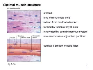
Skeletal muscle structure. striated long multinucleate cells extend from tendon to tendon formed by fusion of myoblasts innervated by somatic nervous system one neuromuscular junction per fiber cardiac & smooth muscle later. fig 9-1a. Skeletal muscle structure. fig 9-2.

Skeletal muscle structure
E N D
Presentation Transcript
Skeletal muscle structure striated long multinucleate cells extend from tendon to tendon formed by fusion of myoblasts innervated by somatic nervous system one neuromuscular junction per fiber cardiac & smooth muscle later fig 9-1a
Skeletal muscle structure fig 9-2
Skeletal muscle structure fig 9-2
Skeletal muscle structure fig 9-3 Don’t bother with: I band, A band, H zone, M line
Skeletal muscle structure 6 thin filaments around each thick 3 thick filaments around each thin fig 9-4
Neuromuscular junction fig 9-14a
Neuromuscular junction fig 9-14b
Generation of motor end plate potential (notes) action potential in somatic motor neuron depolarization of axon terminal, opening of voltage gated Ca++ channels Ca++ enters cell & activates fusion of AcCh vesicles with docking sites AcCh released into synaptic cleft AcCh binds to non-specific ligand gated cation channels in motor end plate opening of channels; Na+ influx greater than K+ efflux motor end plate potential occurs (EPSP) & spreads to edge of plate edge of motor end plate acts like initial segment of axon terminal voltage gated Na+ & K+ channels generate action potential in muscle note: motor nerve action potential always generates muscle action potential Relaxation: AcCh release ends; acetylcholinesterase hydrolyses AcCh; choline transported back into axon terminal
Structure of sarcoplasmic reticulum fig 9-11a
Structure of sarcoplasmic reticulum fig 9-11b
Ca++ release from sarcoplasmic reticulum (s.r.) fig 9-15 cropped action potential spreads across muscle membrane and down T tubules depolarization sensed by dihydropyridine (DHP) receptor in T tubule wall DHP receptor opens ryanodine receptor & its Ca++ channel in s.r. wall Ca++ released into cytosol; subsequently returned to s.r. by Ca++ ATPase
Interaction of thick and thin filaments fig 9-07a Myosin cross bridges bind to sites on actin (when exposed)
Myosin structure fig 9-07b Heavy chains (paired): tail, hinge & cross bridge Light chains (2 pairs): involved in ATPase activity & regulation
Ca++ binds to troponin fig 9-12 Ca++ binds to troponin which causes tropomyosin to move to side exposed sites on actin bind/release myosin cross bridges
Troponin function: low Ca++ fig 9-9a in absence of Ca++: troponin holds tropomyosin against cross-bridge binding site on actin
Troponin function: high Ca++ fig 9-9b in presence of Ca++: troponin moves tropomyosin away from cross-bridge binding site on actin
Cross bridge cycling (notes) Resting state [Ca++], X-bridge binding site covered X-bridge energized (A + *MADPPi) Ca++ release from s.r. [Ca++] exposes X-bridge binding site on actin energized X-bridge binds to actin, ADP & Pi released (step 1) X-bridge “uncocks” as thick filament slides past thin filament (AM) (step 2) ATP gets involved ATP binds to myosin, releasing actin binding (MATP + A) (step 3) X-bridge is energized (cocked) MATP *MADPPi (step 4) Cycling continues until [Ca++] falls
Muscle relaxation action potentials in motor nerve cease AcCh in synaptic cleft hydrolyzed by acetylcholinesterase action potentials in muscle fiber cease Ca++ pumped back into sarcoplasmic reticulum troponin moves tropomyosin to cover X-bridge binding sites myosin remains in *MADPPi form antagonistic muscle extends relaxed muscle
Muscle fiber contraction fig 9-10 This is the response of a single muscle fiber to a single action potential