Exercise and Endothelium
Exercise and Endothelium. دکتر گائینی مجید قلی پور. Exercise and Endothelium. هدف این فصل از کتاب بررسی اجمالی مشارکت اندوتلیوم در روند های بیولوژیکی و در ادامه، تمرکز روی اثر متقابل اعمال این سلول ها و تمرین است. Endothelium.

Exercise and Endothelium
E N D
Presentation Transcript
Exercise and Endothelium دکتر گائینی مجید قلی پور
Exercise and Endothelium • هدف این فصل از کتاب بررسی اجمالی مشارکت اندوتلیوم در روند های بیولوژیکی و در ادامه، تمرکز روی اثر متقابل اعمال این سلول ها و تمرین است.
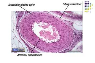
Endothelium • برای مدت زیادی اندوتلیوم به عنوان یک غشاء ساده مورد توجه بوده که روی سیستم گردش خون (عروق) را می پوشاند و عمل اصلی آن حفظ جریان خون و نفوذپذیری دیواره عروق است. • بعداً به عنوان یک اندام پویا که ترشحات حیاتی، متابولیکی، سنتزی و همچنین اعمال ایمنی را انجام می دهد، در نظر گرفته شد. • اندوتلیوم از نظر ساختاری شامل یک لایه مسطح از سلول های بهم متصل شده است که جریان خون را تنظیم نموده و بین داخل عروق و فشای بین سلولی به عنوان یک سد عمل نموده و انتقال مواد غذایی و ملکول های فعال بیولوژیکی مختلف، چسبندگی و عبور سلول های خون به درون بافتها و کنترل هموستاز را تنظیم می نماید. • این اندام از 1013×6 تا 1 اندوتلیوم به وزن تقریبی 1 کیلوگرم تشکیل شده که سطحی حدود 1 تا 7 متر مربع را می پوشاند.
Endothelium Cell Function • در بین وظایف اصلی اندام خودمختار اندوتلیوم، موارد زیر بویژه در مورد فعالیت بدنی از همه جالب تر است. • Regulation of vascular tone. • The induction of alteration in vascular structure. • The interaction with blood cells.
Regulation of vascular tone • اندوتلیوم یک نقش اساسی در تنظیم جریان و فشار خون دارد. • از این جهت تحت کنترل عوامل موجود در موضع خود و همچنین گردش خون سیستمیک می باشد که نتیجه آن تعادل بین افزایش و کاهش قطر عروق است. • عوامل موضعی: • تنظیم خودکار مقدار سفت بودن عروق، به ویژه در پاسخ به محرک های مکانیکی حادث می شود. • دو نیرویی که در پاسخ به فعالیت ورزشی و در نتیجه افزایش جریان خون بر روی عروق خونی عمل می کنند عبارتند از: • 1- کشش ضربانی Pulsatile stretch • 2- فشاری که عروق به خون وارد می نماید Shear stress
Regulation of vascular tone • اثراتی که باعث ایجاد shear stress می شوند عبارتند از : • 1- Vasodilators: such as Nitric axide (NO) and Prostacyclin (PGl2) • 2-Vasoconstrictors: such as endothelin (ET) and platelet factor (PAF). • آزاد شدن این مواد شیمیایی توسط گیرنده های ویژه موضعی در سلول های عروقی تنظیم می شود که از طریق متابولیسم سریع و بر اساس بیان ژن حادث می گردد. • آزاد شدن مستمر NO توسط سلول های اپیتلیوم توسط تعدادی از مواد شیمیایی خارج سلولی و تحریک فیزیکی تعدیل می شود. • دیگر مواد (PGl2,ET and PAF) مقدمتاً در پاسخ به تغییرات محیط خارجی سنتز می شوند.
Regulation of vascular tone • NO اولین بار در سال 1980به عنوان عامل شل کننده اندوتلیوم بیان گردید. Endothelium-drived relaxing factor (EDRF) • NO از اکسیده شدن ال آرژنین به سیترولین توسط NO سنتتاز تشکیل می شود. 3 نوع NO سنتتاز وجود دارد: • 1- NOSI(nNOS) در بافت عصبی. • 2- NOSII(iNOS) در بسیاری از بافت ها لیکن به ندرت. • 3- NOSIII(eNOS) اساساً در سلول های اندوتلیوم و همچنین در عظلات قلبی و پلاکت ها.
Vasodilatory Agonists • eNOS در سلول های اندوتلیوم به طور قوی فعال است. • eNOS با افزایشCa2+ داخل سلولی تحریک می گردد. ضمناً اتصال برخی از مواد (مانند thrombin، bradykiniadenosine 5-disphosphate، substance P) به رسپتورهای غشاء می تواند باعث افزایش Ca2+ داخل سلولی و در نتیجه تحریک eNOS شود. • NO اثرات شل کننده گی خود را بر روی عضلات صاف عروق با فعال کردن چرخه soluble guanylate و تولید cGMP اعمال می کند. • همچنین ممکن است NO باعث تحریک کانال های پتاسیمی وابسته به Ca2+شود که منجر به هایپرپولاریزاسیون و شل شدن عضلات صاف عروق می گردد.
Vasodilatory Agonists • مسیر وابسته به Ca2+ باعث افزایشCa2+ داخل سلولی و فعال شدن فسفو لیپاز c می گردد. • مسیر مستقل از Ca2+به فعالیتsodium-hydrogen exchanger و فعال شدن پروتئین کیناز c و تیروئین کیناز نیازمند است. • افزایش قطر عروق در اثر جریان خون نیازمند آزاد شدن عوامل زیر می باشد: • ATP • substances P • Prostacyclin • Endothelium-drived hyperpolarization factor (EDHF)
Vasodilatory Agonists • ATP and Substance P which are release from endothelium cells by shear stress, stimulate specific receptors and cause the release of NO. • A burst of Prostacyclin produced by endothelium cells could be observed after a sudden increase of shear stress. • EDHF is a less well described product of endothelium cells. Stimulation with muscarine agonists leads to a release of EDHF causing a transient hyperpolarization of the cell membrane.
Vasoconstrictory Agonists • مهمترین موادی که باعث انقباض عروق می شوند عبارتند از: Endothelin-1 (ET-1) and Angiotensin II (AT2) • آنژیو تانسین II بوسیله سلول های اندوتلیوم ترشح نمی شود لیکن توسط Engiotensin converting enzyme (ACE) ، در سطح اندوتلیوم تولید می شود.
Vasoconstrictory Agonists • تا این تاریخET-1 یکی از قویترین پپتیدهای شناخته شده در انقباض عروق می باشد که در اثر بیان ژن preproendothelin-1 که پیش ساز ET-1 است، تشکیل می شود. • ET-1 با اتصال به رسپتور ET-A که به وفور در سلول های عضلات صاف عروق وجود دارد، باعث افزایش غلظتCa2+ داخل سلولی و در نتیجه افزایش تون در عضلات صاف عروق می گردد. • NO بلافاصله، با شتاب دادن به ذخیره شدن مجدد Ca2+ باعث برگشت تون عضله به سطح قبلی می گردد. • سطح فیزیولوژیکیshear stress ، به طور موقتی باعث افزایش up-regulation بیان ET-1mRNA شده که به دنبال آن سرکوب می گردد که در سلول های اندوتلیوم انسان با Low shear stress افزایش می یابد لیکن با High shear stress مهار می گردد.
Vasoconstrictory Agonists • Angiotansin II همانند renin-angiotensin، یک منقبض کننده عروق قوی می باشد که با NO در اندوتلیوم واکنش متقابل دارد. • Angiotensin converting enzyme (ACE) بر روی سطح اندوتلیوم نه تنها مسئول تبدیل آنژیوتانسین I به آنژیوتانسین II می شود بلکه باعث غیر فعال شدن bradykinin و کاهش ترشح NO وابسته به آن می گردد. تنظیم تون عضلات عروق با استفاده از مواد تنگ و گشاد کننده Endothelium-dependent regulation of vascluar tone نام دارد.
Endothelium-independent regulation of vascular tone کشش مکانیکی سبب عمل این مکانیزم می شود. • کشش منجر به فعال شدن کانال یونی غیر انتخابی وابسته به کشش بر روی غشاء پلاسمایی می شود که نتیجه آن ورود کلسیم به داخل سلول و دپولاریزاسیون آن است. • nonselective stretch-activation cation (SAC) channel • دپولاریزاسیون به نوبه خود کانال کلسیمی وابسته به ولتاژ را فعال می کند که منجر به ورود کلسیم (علاوه بر ورود کلسیم از SAC) به داخل سلول می گردد. • ورود کلسیم از هر دو کانال منجر به انقباض عضله سفید می گردد. • در مقابل، ورود کلیسیم و دپولاریزاسیون غشاء، باعث فعال شدن کانال پتاسیمی وابسته به ولتاژ و خروج پتاسیم از سلول و در نتیجه هایپرپلاریزه شدن سلول می شود. • این عمل یک مکانیزم بازخورد منفی برای محدود نمودن دپولاریزاسیون و انقباض عضله صاف است.
Endothelium-independent regulation of vascular tone Stretch Activation of cation channelin the plasma membrane Depolarization voltage-gated Ca2+ channel Ca2+ influx Smooth muscle contraction K+ channel Voltage-dependent K+ outflux Hyperpolarization
Endothelium-independent regulation of vascular tone Stretch Activation of cation channelin the plasma membrane Depolarization voltage-gated Ca2+ channel Ca2+ influx Smooth muscle relaxation K+ channel Voltage-dependent K+ outflux Hyperpolarization
Vascular Control in Skeletal Muscle atRest and During Exercise • Endothelium-derived NO is a significant regulator of basal vascular tone in resting muscle. • It’s contribution to exercise-induced vasorelaxation in active skeletal muscle is less clear. No may be responsible for the initial (1 min) hyperemic response mediating the dilatation of first- and second-order arteries, but not third- order arteries. • Blockage of endothelium-derived NO did not alter the exercise-induced hyperemia in skeletal muscle of cat or rabbit.
Vascular Control in Skeletal Muscle atRest and During Exercise • In human, interabrachial infusion of NO inhibitors produced a decrease of forearm blood flow at rest as well as during rhythmic and static exercise. • Because of the same magnitude of reduction in blood flow at rest and during exercise, these finding suggest a lack of additional NO-mediated control of muscle perfusion during exercise. • Whether other endothelium-derived vasodilators are involved remains to be investigated.
Flow-Induced Alterationsin Vascular Structure • عواملی که در تنظیم تون عروق در گیر هستند، مسئول تغییراتی در ساختار عروق هم هستند. • به طور مشابه، مسیری که بر اثر جریان خون باعث شل شدن عروق می شود، در بیان تغییر شکل growth factor-beta هم می گردد. • از طرف دیگر ET-1 (منقبض کننده عروق) یک ماده قوی میتوژن است که می تواند بوسیله دیگر عواملی که رشد را تحریک می کنند مانند angiotensin II، platelet-derived growth factor-beta و insulin پرتوان گردد.
Flow-Induced Alterationsin Vascular Structure • عروق در پاسخ به تغییرات طولانی مدت جریان خون توانایی تغییر وضع دادن عمده ای دارند. تمرینات استقامتی در حیوانات باعث افزایش قطر شریان کاروتید و کرونری می شود که مستقل از واکنش انتهای این اندام ها می باشد. • در مقابل، کاهش طولانی مدت جریان خون باعث کاهش پاسخ تغییرات ساختاری در دیواره عروق می شود.
Flow-Induced Alterationsin Vascular Structure • Identified mediators involved in cell growth, extracellular matrix production, and proteolysis are: 1- ET-1. 2- NO. 3-Plasminogen-drived growth factor (PDGF-beta). 4- prostacyclin. 5- Transforming growth factor-beta (TGF-beta). 6- Tissue plasminogen activator (t-PA).
Flow-Induced Effect on Hemostasis/Thrombolysis/Fibrinolysis • NO ضمن افزایش قطر عروق، اثرات چشم گیری در هموستاز دارد. • NO تولید شده توسط NOSIII در پلاکت ها، چسبندگی، فعال شدن، تراوش و تجمع خود پلاکت ها را مهار می کند و تجزیه آن ها را بیشتر می نماید. • مکانیزمی که باعث این اثرات می شود تا حدودی به cGMP وابسته است.
Flow-Induced Effect on Hemostasis/Thrombolysis/Fibrinolysis • با توجه به فیبرینولیز، نشان داده شده کهshear stress یک محرک قوی در بیان فعال کننده پلاسمینوژن بافتی و تولید پروتئین در خارج از بدن است. • در جایی که افزایش سیتوپلاسمی Ca2+ باعث رهایی t-PA می شود، این فرضیه شکل گرفته که افزایش Ca2+ در اثر فعالیت ورزشی می تواند باعث افزایش ظرفیت آزاد شدن t-PA شود. • NO، اثر لوکوسیت ها را بر دیواره عروق مهار می نماید. این عمل را یا با جلوگیری از فعال شدن لوکوسیت ها و یا با ایجاد ملکول های چسبنده اعمال می نماید.
Effect of Physical Training onVascular Reactivity • با تأکید بر نقش مهم ورزش در درمان تنوس زیاد عضله، فعالیت بدنی به عنوان یک تعدیل کننده فشار خون شناخته شده است. • اسناد تحقیقات اخیر بیان می دارد که دیواره عروق، محلی است که تمرین بر روی آن اثر کرده و نسبت به فعالیت های بدنی طولانی مدت سازگار می شود. • گذشته از اثرات لحظه ای افزایش جریان خون، اسناد مدارکی در دست است که افزایش طولانی مدت جریان خون (همانگونه که در تمرینات ورزشی بوجود می آید) باعث ایجاد اثرات سودمندی بر روی واکنش پذیری عروق می شود. • این سازگاری به عنوان یک بهبود همه جانبه عروق در افزایش قطر و کاهش انقباض، نسبت به عوامل شیمیایی می باشد.
Receptor/Channel/Signaling Level • The effect of training can be seen in enhancement: • In the expression of receptors • In the contribution of channels • On the signaling level.
Transcriptional Level • The training-induced long-term increase in blood flow modulates the expression of NO synthase. • In cultured endothelium cells expose to long-term shear stress is NO synthesae up-regulated.
Factors of Influence • There are several factors that are important for the linkage between exercise and vascular adaptation. • Magnitude and nature of flow. • Vessel size. • Frequency of exercise. • Type of exercise. • Time point of exercise training adaptation.
The Effect of detraining • In a study the plasma concentration of NO increased significantly while the plasma level of ET-1 decreased significantly at the end of eight-week aerobic exercise training (bicycle ergometer) and lasted through the fourth week after cessation of exercise training. • These changes returned to the pre-exercise level after the eighth week. • There was a negative correlation between plasma NO and ET-1 concentration.
Age • Ageing is associated with a progressive decline in endothelium-dependent dilatation. • In a study, using ultrasound technique, showed that the vasodilatator response, in male subject younger than 40 years was %70 higher than in older ones. • Such differences were less pronounced when female subject were examined. However, another study demonstrated a progressive impairment in endothelium function in the ageing process for both genders. An explanation for the contradictory finding might be that the process occurs in men and women at different age.
Age • Several study showed that aerobic exercise training can restore the age-related loss of endothelium-dependent vasorelaxation in old men and women even in previously sedentary ones.
Conclusion • Finally, it is important to keep in mind that all the findings presented in this chapter have many limitation. • Most conclusion drawn from these studies are based on either in vitro or animal models, with only a few derived from human studies. • The others limitation are: • Broad variation in investigation techniques. • Endothelium of different vessels, which differ in size and function. • Different exercise type (intensity, duration).
Conclusion • Although data are conflicting, there is an overall trend toward an improvement of endothelium function caused by exercise. • Beside on the presumption that endothelium dysfunction is systemic nature, one might assume that regular, especially aerobic, exercise produces favorable global endothelial adaptations. • To complete the inchoate puzzle of existing data, further investigation is necessary.