Tissue
Tissue. Chapter 4. Link. Tissues. Tissue: 4 Primary Tissue Types Epithelial Connective Muscle Nervous. http://www.stegen.k12.mo.us/ tchrpges / sghs / ksulkowski /images/20_Simple_Columnar_Epithelial_Tissue.jpg. Match Tissue Type to Function. Epithelial Connective Nervous Muscle.

Tissue
E N D
Presentation Transcript
Tissue Chapter 4 Link
Tissues • Tissue: • 4 Primary Tissue Types • Epithelial • Connective • Muscle • Nervous http://www.stegen.k12.mo.us/tchrpges/sghs/ksulkowski/images/20_Simple_Columnar_Epithelial_Tissue.jpg
Match Tissue Type to Function • Epithelial • Connective • Nervous • Muscle • Supports, protects, binds other tissues together • Internal communication • Contracts to cause movement • Forms boundaries between different environments, protects, secretes, absorbs, filters
Epithelial Tissue (Epithelium) • Two main types (by location): • Covering and lining epithelium • Glandular epithelium Forms boundaries b/w different environments http://www.bio.davidson.edu/people/kabernd/BerndCV/Lab/EpithelialInfoWeb/goblet%20cells%20.jpg
Functions of Epithelial Tissue • Protection • Absorption • Filtration • Excretion • Secretion • Sensory reception
Characteristics of Epithelial Tissue • Cells have polarity • Are composed of closely packed cells • Supported by a connective tissue reticular lamina (under the basal lamina) • Avascular but innervated • High rate of regeneration
Classification of Epithelia • Ask two questions: • How many layers? 1 = simple epithelium >1 = stratified epithelium
Classification of Epithelia • What type of cell? • Squamous • Cuboidal • Columnar Note: if stratified, name according to apical layer of cells!
Overview of Epithelial Tissues • For each of the following types of epithelia, note: • Description • Function • Location
Simple Epithelia • Single cell layer (usually very thin) • Concerned with: • Absorption • Secretion • Filtration • NOT concerned with: protection • Simple squamous, simple cuboidal, simple columnar, pseudo stratified columnar
Simple Squamous Epithelium • Description • Function • Location Photomicrograph: Simple squamous epithelium forming part of the alveolar (air sac) walls (125x). Note: ENDOTHELIUM AND MESOTHELIUM
Simple Cuboidal Epithelium (b) Simple cuboidal epithelium • Description • Function • Location Photomicrograph: Simple cuboidal epithelium in kidney tubules (430x).
Simple Columnar Epithelium (c) Simple columnar epithelium • Description • Function • Location Photomicrograph: Simple columnar epithelium of the stomach mucosa (860X).
Pseudostratified Columnar Epithelium (c) Simple columnar epithelium • Description • Function • Location Photomicrograph:Pseudostratified ciliated columnar epithelium lining the human trachea (570x).
Stratified Epithelium • 2+ cell layers • Regenerate from below • More durable than simple epithelia • Major role: Protection
Stratified Squamous Epithelium • Description • Function • Location Photomicrograph: Stratified squamous epithelium lining the esophagus (285x).
Stratified Cuboidal Epithelium • Description • Function • Location
Stratified Columnar Epithelium • Description • Function • Location http://www.sciencephoto.com/image/115414/large/C0051252-Stratified_columnar_epithelium,_urethra-SPL.jpg
Transitional Epithelium • Description • Function • Location Photomicrograph: Transitional epithelium lining the urinary bladder, relaxed state (360X); note the bulbous, or rounded, appearance of the cells at the surface; these cells flatten and become elongated when the bladder is filled with urine.
Glandular Epithelia • Gland: one or more cells that secretes and aqueous fluid • Classified by: • Site of product release • Endocrine • Exocrine • Relative number of cells forming the gland • Unicellular • Multicellular
More numerous! Glands Endocrine Exocrine Secrete products into ducts Secretions released onto body surfaces (skin) or into body cavities Examples: mucous, sweat, oil, and salivary glands • Ductless glands • Secrete hormones that travel through lymph or blood to target organs • Examples: Thyroid Gland, Pituitary Gland • Covered in Ch. 16
Unicellular Exocrine Glands • Goblet cell and Mucous cell • Mucin -> mucous
Multicellular Exocrine Glands • Composed of a duct and a secretory unit • Classified according to: • Duct type • Simple • Compound • Structure of secretory units • tubular • alveolar • tubuloalveolar
Simple duct structure (duct does not branch) Compound duct structure (duct branches) Tubular secretory structure Simple tubular Simple branched tubular Compound tubular Example Intestinal glands Example Stomach (gastric) glands Example Duodenal glands of small intestine Alveolar secretory structure Simple alveolar Simple branched alveolar Compound alveolar Compound tubuloalveolar Example No important example in humans Example Sebaceous (oil) glands Example Mammary glands Example Salivary glands Surface epithelium Duct Secretory epithelium Figure 4.5
Modes of Secretion Merocrine Holocrine Products are secreted by rupture of gland cells sebaceous (oil) glands • Products are secreted by exocytosis • pancreas, sweat and salivary glands
Connective Tissue • Most abundant and widely distributed tissue type • Four main classes • Connective Tissue Proper • Cartilage • Bone Tissue • Blood See Table 4.1
Major Functions of Connective Tissue • Binding and Support • Protection • Insulation • Stores reserve fuel • Transports
Characteristics of Connective Tissue • Connective tissues have: • Mesenchyme as their common tissue of origin • Varying degrees of vascularity • Cells separated by nonliving extracellular matrix (ground substance and fibers) • 3 Structural Elements • Ground substance • Fibers • Cells
Structural Elements of Connective Tissue • Ground substance • Medium through which solutes diffuse between blood capillaries and cells • Components: • Interstitial fluid • Adhesion proteins (“glue”) • Proteoglycans • Protein core + large polysaccharides • Trap water -> viscosity
Structural Elements of Connective Tissue • Connective Tissue Fibers • Collagen (white fibers) • Strongest and most abundant type • Provides high tensile strength • Elastic (yellow fibers) • Networks of long, thin, elastin fibers that allow for stretch/recoil • Reticular • Short, fine, highly branched collagenous fibers
Structural Elements of Connective Tissue • Cells (see table 4.1) • Mitotically active and secretory cells = “blasts” • Fibroblasts, chondroblasts, osteoblasts, hematopoietic stem cells • Mature cells = “cytes” • Chondrocytes, osteocytes • Other cell types • Fat cells, white blood cells, mast cells, and macrophages
Cell types Extracellular matrix Ground substance Fibers • Collagen fiber • Elastic fiber • Reticular fiber Macrophage Fibroblast Lymphocyte Fat cell Capillary Mast cell Neutrophil Figure 4.7
Connective Tissue: Embryonic • Mesenchyme—embryonic connective tissue • Gives rise to all other connective tissues • Gel-like ground substance with fibers and star-shaped mesenchymal cells
Types: Loose connective tissue Areolar Adipose Reticular Dense connective tissue Dense regular Dense irregular Elastic Connective Tissue Proper
CONNECTIVE TISSUE PROPER Loose Connective: Areolar • Description • Function • Location Photomicrograph: Areolar connective tissue, a soft packaging tissue of the body (300x).
CONNECTIVE TISSUE PROPER Loose Connective: Adipose • Description • Function • Location Photomicrograph: Adipose tissue from the subcutaneous layer under the skin (350x).
CONNECTIVE TISSUE PROPER Loose Connective: Reticular • Description • Function • Location Photomicrograph: Dark-staining network of reticular connective tissue fibers forming the internal skeleton of the spleen (350x).
CONNECTIVE TISSUE PROPER Dense Connective: Dense Regular • Description • Function • Location Photomicrograph: Dense regular connective tissue from a tendon (500x).
CONNECTIVE TISSUE PROPER Dense Connective: Dense Irregular • Description • Function • Location Photomicrograph: Dense irregular connective tissue from the dermis of the skin (400x).
CONNECTIVE TISSUE PROPER Dense Connective: Elastic • Description • Function • Location Photomicrograph: Elastic connective tissue in the wall of the aorta (250x).
Connective Tissue: Cartilage • Stands up to both compression and tension • No nerve fibers, avascular • 80% water • Chondroblasts– produce new matrix • Chondrocytes – mature cartilage cells • Found in small groups in lacunae
CARTILAGE Hyaline Cartilage • Description • Function • Location Photomicrograph: Hyaline cartilage from thetrachea (750x).
CARTILAGE Elastic Cartilage • Description • Function • Location Photomicrograph: Elastic cartilage fromthe human ear pinna; forms the flexibleskeleton of the ear (800x).
CARTILAGE Fibrocartilage • Description • Function • Location Photomicrograph: Fibrocartilage of an intervertebral disc (125x). Special staining produced the blue color seen.
Connective Tissue: Bone • Description • Function • Location Photomicrograph: Cross-sectional viewof bone (125x).
Connective Tissue: Blood • Description • Function • Location Photomicrograph: Smear of human blood (1860x); twowhite blood cells (neutrophil in upper left and lymphocytein lower right) are seen surrounded by red blood cells.
Nervous Tissue • Description • Function • Location Photomicrograph: Neurons (350x)
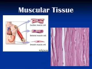
Muscle Tissue • Highly cellular, well vascularized • Movement • Types • Skeletal • Cardiac • Smooth
MUSCLE TISSUE Skeletal Muscle • Description • Function • Location Photomicrograph: Skeletal muscle (approx. 460x). Notice the obvious banding pattern and the fact that these large cells are multinucleate.
MUSCLE TISSUE Cardiac Muscle • Description • Function • Location Photomicrograph: Cardiac muscle (500X);notice the striations, branching of cells, andthe intercalated discs.