Effects of Sodium Iodide on Peptide Stability: Computational Analysis
Investigating sodium iodide's impact on the helical stability of an mainly-alanine peptide using molecular dynamics simulations and experimental techniques.

Effects of Sodium Iodide on Peptide Stability: Computational Analysis
E N D
Presentation Transcript
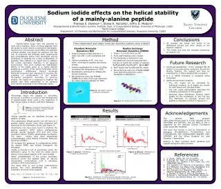
1” outside border 42” wide Logo should be of your sponsor (PSC, Pitt, CMU, Duquesne) Sodium iodide effects on the helical stability of a mainly-alanine peptideTheresa E. Downey1,2, Eliana K. Asciutto3, Jeffry D. Madura31Bioengineering & Bioinformatics Summer Institute, Dept. of Computational Biology, University of Pittsburgh, 152602Saint Vincent College3Department of Chemistry and Biochemistry for Computational Sciences, Duquesne University, 15282 Logo for your home school 0.5” inside borders Abstract Method Conclusions Two independent and unique molecular dynamics systems were created On average the iodide ions prefer to be distributed around and more closely to the arginine residues. The iodide ions have the strongest preference for arginine 14. Peptide-based drugs have the potential to treat many diseases. Some α-helical peptides have the ability to enter cells by crossing the lipid bilayer. However, the environment could affect the peptide’s α-helical stability and hinder its entrance into the cell. To overcome this difficulty both experimental and computational teams have developed various techniques to stabilize the α-helix configuration of peptides. One method is to select ions to stabilize the α-helix secondary structure in an aqueous environment. This work studied the stabilization effects of the iodide anion on a mainly alanine peptide as a portion of the entire Hofmeister series investigation. Additionally, Replica Exchange Molecular Dynamics was employed to increase the sampling of configurations of the system throughout simulation. According to previous studies, the iodide ion is expected to stabilize the peptide less than perchlorate in an aqueous environment according to the Hofmeister series. Standard Molecular Dynamics (MD) Replica Exchange Molecular Dynamics (REMD) • Steps 1-2 are the same as MD. • Forty-eight replicas were created. • The temperature distribution was calculated with the following geometric function to match the number of replicas: Tj=T1(1+((T2-T1)/T1))(j-1) (j=1-48)5 • Each replica was equilibrated for 200ps • MD performed using the FF99SB AMBER force field throughout the entire temperature distribution. • Alanine-peptide initially placed in a water box with I- and neutralized with Na+. • System consisting of AP, ions, and water minimized to stabilze decreasing energy. • System equilibrated for a total of 200ps increasing the temperature to 300K. • Equilibration continued for 200ps/until density stabilizes. • MD performed using the FF99SB AMBER force field, which was optimized for mainly-alanine peptides6 at 300K. Future Research • Continued equilibration of the standard MD AP system is currently being conducted. Should the density stabilize, the system will then be submitted for a 200ns standard MD simulation. • A 1 μs REMD simulation is presently being conducted. • Additional simulation analysis: • The {Ψ} angles will be calculated for individual residues, as Φ angles are similar for both folded and unfolded states. • The {Ψ} angle appropriately describes the unfolding mechanism in this peptide.5 • Generate radial distribution function (RDF) graphs for the simulations to see how the ions distribute, on average, around the peptide comparable to those in Figures 5-7. Feel Free to adjust sizes of any rounded rectangles to fit your needs Introduction Figure 3. AP system used for continued equilibration 36” tall • Knowledge about the stability in secondary structures of proteins is critical in the development of peptide-based drugs used in the treatment of HIV, various types of cancer, and other diseases.1 • Helical peptides can be stabilized through the addition of salts. • The stabilization effects specific ions will have on a peptide and/or protein have been predicted and described by the Hofmeister Series (see Figure 2). • Perchlorate ions have been found to stabilize the alanine peptide’s (AP’s) helical configurations through circular dicroism (CD) and ultra violet resonance Ramen (UVRR) spectroscopy techniques. The same results have been estabilished computationally using Replica Exchange Molecular Dynamics (REMD).3 • To increase the sampling of configurations within the system REMD was utilized throughout the simulation. • Iodide is directly to left of perchlorate within the Hofmeister series. In this work the iodide anions effects on the helical stability of AP were studied. • When destabilization of the α-helix secondary structure occurs the peptide takes on varying intermediate conformations until it ultimately unfolds to PPII conformations.4,5 • The more PPII, β, and αL-helix conformations present, the more destabilized the α-helix secondary structure of the peptide. Figure 1. Example of peptide-based drug developed by Gregory L. Verdine, Harvard University chemical biologist.2 Figure 4. AP system used for the REMD simulation http://pubs.acs.org/cen/coverstory/86/8622cover.html Results Acknowledgements The national BBSI program (http://bbsi.eeicom.com) is a joint initiative of the NIH-NIBIB and NSF-EEC, and the BBSI @ Pitt is supported by the National Science Foundation under Grant EEC-0234002. This work was funded in part by Department of Education (P116Z040100, P116Z050331, and P116Z080180), PSC (MCB060059P, AAB/PSC CHE-030008P) and the Silicon Graphics Inc. and Gaussian Corporations. Kathryn K. Myer provided much guidance and support during the entire process. Colors turn out a bit different on paper than the screen. See Jason for samples. Figure 5 (above). Distribution of iodide around alanine Figure 6 (above). Distribution of iodide around arginine References • C. Drahl, C&EN. 86, 18-23 (2008). • F. Bernal; A.F. Tyler; S.J. Korsmeyer; L.D. Walensky; G.L. Verdine, J. Am. Chem. Soc. 129, 2456-2457 (2007). • E.K. Asciutto; J.D. Madura, In Preparation. 2008. • A.V. Mikhonin; S.V. Bykov; N.S. Myshakina; S.A. Asher, J. Phys. Chem. 110, 1935 (2006). • E.K. Asciutto; A.V. Mikhonin; S.A. Asher; J.D. Madura, Biochemistry. 47, 2046 (2007). • V. Hornak; R. Abel; A. Okur; B. Strockbine; A. Roitberg; C. Simmerling, PROTEINS: Structure, Function, and Bioinformatics. 65, 714 (2006). H2PO4- > SO42- > F- > CH3COO− > Cl- > Br- > NO3- > I- > ClO4- > SCN- Figure 2. The Hofmeister Series Figure 7. Distribution of iodide around arginine 9 (red line), around arginine 14 (green line), and around arginine 19 (blue line)