Download
1 / 31
310 likes | 461 Vues
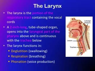
Anatomy of Larynx. General principles of development. The development of the larynx can be divided into prenatal and postnatal stages. At birth, the larynx is located high in the neck between the C1 and C4 vertebrae, allowing concurrent breathing or vocalization and deglutition.

E N D
General principles of development • The development of the larynx can be divided into prenatal and postnatal stages. • At birth, the larynx is located high in the neck between the C1 and C4 vertebrae, allowing concurrent breathing or vocalization and deglutition. • By age 2 years, the larynx descends inferiorly; by age 6 years, it reaches the adult position between C4 and C7 vertebrae. This new position provides a greater range of phonation (because of the wider supraglottic pharynx) at the expense of losing this separation of function, i.e., deglutition and breathing.
The larynx develops from the endodermal lining and the adjacent mesenchyme of the foregut between the fourth and sixth branchial arches. • At 20 days' gestation, the foregut is first identifiable with a ventral laryngotracheal groove. It continues to deepen until its lateral edges fuse. • Trachea becomes separated from the esophagus by the tracheoesophageal septum with a persistent slit like opening into the pharynx • This fusion occurs in the caudal-to-cranial direction, and incomplete fusion results in development of persistent communication between the larynx or trachea and the esophagus
The main changes occurring in the larynx postnatally are a change in the axis, luminal shape, length, and proportional growth of the laryngeal elements. • The larynx grows rapidly during the first 3 years of life, while the arytenoids remain approximately the same size. • Beginning at age 18-24 months, the larynx descends in the neck to achieve its final position at vertebrae C4-C7 by age 6 years. • The larynx elongates as the hyoid, thyroid, and cricoid cartilages separate from each other • The cricoid cartilage continues to develop during the first decade of life, gradually changing from a funnel shape to a wider adult lumen; therefore, it is no longer the narrowest portion of the upper airway.
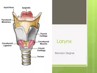
Thyroid Cartilage • Shied shaped, open posteriorly, angulated anteriorly • Angulation more acute in males • Its function is to shield larynx from injury and provide an attachment to vocal cords
Cricoid Cartilage • Signet ring shaped • Stronger than thyroid cartilage. • Lamina – 2 to 3 cm from above downwards, considerably broader than anterior arch.
Important from structural & functional point of view • Base for entire larynx • Support to arytenoid • Attachment to intrinsic muscles • Only part of cartilagenous framework that forms continuous 360 degree ring • Once injured or strictured , difficult to resect while preserving laryngeal function
Epiglottis • Thin leaf shaped fibro-cartilage, situated in midline • Upper free end broad & rounded, projects up behind base of tongue • Narrow base called pitiole • This attachment forms lower limit of pre-epiglottis space
Half of epiglottis projects above hyoid • This part has a laryngeal and lingual surfaces
Infrahyoid portion has no free anterior surface • Forms posterior wall of PES • Epiglottic cartilage contains many pits filled with mucous glands • Little barrier between infrahyoid portion and PES
Arytenoids • Paired cartilages, pyramidal in shape • Base articulated with cricoid • PCA & LCA muscles attach on muscular process • Anterior angle elongated into vocal process which receives insertion of vocal ligament
Supraglottis • Consists of ventricles, false cords, laryngeal surface of epiglottis, aryepiglottic folds and the mucosal expanse. • Posterior tapering shape reduces area of mucosa in posterior region • So majority of SG tumors are epiglottic
Glottis • Consists of true cords, anterior commissure and posterior commissure • Narrow triangular space between the true cords is called rima glottis • Anterior 2/3 is membranous • Posterior third consists of vocal processes of arytenoids • Posterior 1/3 of cords and covering mucosa are called posterior commissure
Sub-glottis • Begins about 5mm below free margins of VC • Consists of a mobile upper and fixed lower part
Mucosa • Mucosa of glottic and Supraglottic regions is stratified squamous epithelium. • Mucosa of ventricles and sub-glottic regions is pseudo-stratified ciliated epithelium • Supra and sub glottic regions particularly ventricles are rich in submucosal mucous or minor salivary glands while glottis is not.
Nerve Supply: Derived from the Vagus • Superior Laryngeal Nerve -It leaves the vagus nerve high in the neck • Internal -It provides sensation of the glottis and supraglottis, which includes the pharynx, underside of the epiglottis and the larynx above the cords. Remember: SIS-superior internal sensory. • External -It supplies motor function to the cricothyroid muscle which tenses the vocal cords and could cause laryngopasm.
Recurrent Laryngeal Nerve -It provides sensation to the subglottic area which includes the larynx below the vocal cords and upper esophagus. It provides motor function to the intrinsic muscles of the larynx. • It branches from the vagus in the mediastinum and turns back up into the neck. On the right, it travels inferior to the subclavian and loops up, and on the left it travel inferior to the aorta and loops up.
ARTERIAL SUPPLY • Sup. Laryngeal A. from Sup. Thyroid artery • Inf. Laryngeal A. from Inf. Thyroid artery
Pre-Epiglottic Space • Bound sup by hyo-epiglottic ligament, ant by thyrohyoid memb. & thyroid cartilage and posteriorly by epiglottis • Filled with fat and areolar tissue • Continuous with para-glottic space • Cx of laryngeal surface of epiglottis readily spread to PreEpiSpace
Reinke’s Space • Mucosa over the vocal ligament loosely attached to ligaments • Thus there is a submucosal space along most of the length of truer VC
Intrinsic Ligaments of larynx • Quadrangular Membrane • Conus Elasticus