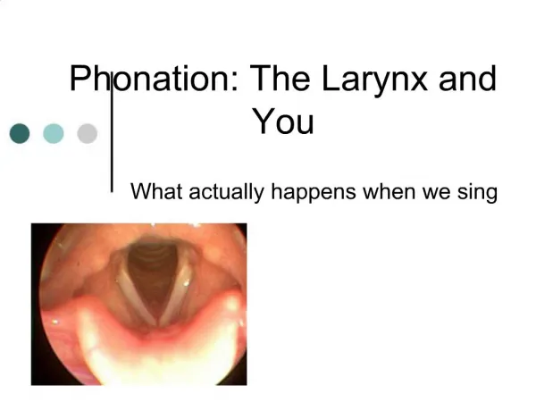
The Larynx
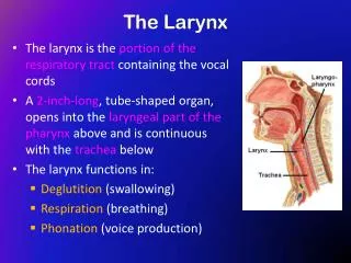
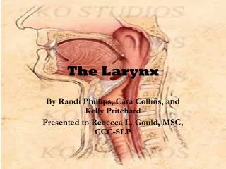
The Larynx. Prof. Dr.Mohammed Hisham Al-Muhtaseb. The Larynx. Extends from the middle of C3 vertebra till the level of the lower border of C6 Continue as Trachea Above it opens into the laryngo-pharynx

The Larynx
E N D
Presentation Transcript
The Larynx Prof. Dr.Mohammed Hisham Al-Muhtaseb
The Larynx • Extends from the middle of C3 vertebra till the level of the lower border of C6 • Continue as Trachea • Above it opens into the laryngo-pharynx • Suspended from the hyoid bone above and attached to the trachea below by membranes and ligaments
Functions • 1. acts as an open valve in respiration • 2. Acts as a closed valve in deglutition • 3. Acts as a partially closed valve in the production of voice • 4. During cough it is first closed and then open suddenly to release compressed air
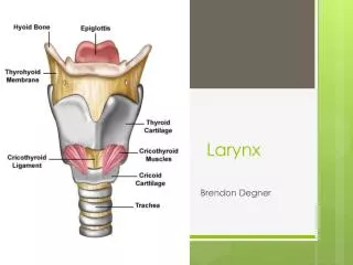
Parts • 1. Cartilage • 2. Mucosa • 3. Ligaments • 4. Muscles
Cartilage • A. Single : Epiglottis Cricoid Thyroid B. Pairs: Arytenoid Cuneiform Corniculate
Cricoid cartilage • The most inferior of the laryngeal cartilages • Completely encircles the airway • Shaped like a 'signet ring' • Broad lamina of cricoid cartilage posterior • Much narrower arch of cricoid cartilage circling anteriorly.
Cricoid cartilage • Posterior surface of the lamina has two oval depressions separated by a ridge • The esophagus is attached to the ridge • Depressions are for attachment of the posterior crico-arytenoid muscles. • Has two articular facets on each side • One facet is on the sloping superolateral surface and articulates with the base of an arytenoid cartilage; • The other facet is on the lateral surface near its base and is for articulation with the inferior horn of the thyroid cartilage
Thyroid cartilage • The largest of the laryngeal cartilages • It is formed by a right and a left lamina • Widely separated posteriorly, but converge and join anteriorly • Posterior margin of each lamina is elongated to form a superior horn and an inferior horn
Thyroid cartilage • Most superior point of the site of fusion between the two laminae is the laryngeal prominence ('Adam's apple') • Angle between the two laminae is more acute in men (90°) than in women (120°) • Superior to the laryngeal prominence, the superior thyroid notch separates the two laminae • Superior thyroid notch and the laryngeal prominence are palpable landmarks in the neck • Less distinct inferior thyroid notch in the midline along the base of the thyroid cartilage.
Thyroid cartilage • The medial surface of the inferior horn has a facet for articulation with the cricoid cartilage; • The superior horn is connected by a ligament to the posterior end of the greater horn of the hyoid bone. • Lateral surface of lamina is marked by a ridge (the oblique line), which curves anteriorly from the base of the superior horn to the inferior margin of the lamina. • Ends of the oblique line are expanded to form superior and inferior thyroid tubercles • The oblique line is a site of attachment for the extrinsic muscles of the larynx (sternothyroid, thyrohyoid, and inferior constrictor).
Epiglottis • Is a 'leaf-shaped' cartilage attached by its stem to the angle of the thyroid cartilage • Projects posterosuperiorly from its attachment to the thyroid cartilage. • The attachment is via the thyro-epiglottic ligament in the midline between the laryngeal prominence and the inferior thyroid notch • The upper margin of the epiglottis is behind the pharyngeal part of the tongue. • The inferior half of the posterior surface of the epiglottis is raised slightly to form an epiglottic tubercle.
Arytenoid cartilages • Two arytenoid cartilages are pyramid-shaped cartilages with three surfaces • Base of arytenoid cartilage and an Apex of arytenoid cartilage • The base of arytenoid cartilage is concave and articulates with the facet on the superolateral surface of the cricoid cartilage; • The apex of arytenoid cartilage articulates with a corniculate cartilage; • The medial surface of each cartilage faces the other;
Arytenoid cartilages • The anterolateral surface has two depressions, separated by a ridge, for muscle (vocalis) and ligament (vestibular ligament) attachment. • The anterior angle of the base of arytenoid cartilage is elongated into a vocal process to which the vocal ligament is attached • The lateral angle is similarly elongated into a muscular process for attachment of the posterior and lateral crico-arytenoid muscles.
Corniculate and Cuneiform • The corniculate cartilages are two small conical cartilages • Bases articulate with the apices of the arytenoid cartilages • Their apices project posteromedially towards each other. • The Cuneiform are two small club-shaped cartilages • Lie anterior to the corniculate cartilages • Suspended in the part of the fibroelastic membrane that attaches the arytenoid the epiglottis.
Extrinsic ligaments • Thyrohyoid membrane • Hyo-epiglottic ligament • Cricotracheal ligament
Thyrohyoid membrane • Tough fibroelastic ligament that spans between the superior margin of the thyroid cartilage below and the hyoid bone • Attached to the thyroid laminae and adjacent anterior margins of the superior horns • Ascends medial to the greater horns and posterior to the body of the hyoid bone to attach to the superior margins of these structures. • An aperture in the lateral part of the thyrohyoid membrane on each side is for the superior laryngeal arteries, nerves, and lymphatics
Thyrohyoid membrane • The posterior borders of the thyrohyoid membrane are thickened to form the lateral thyrohyoid ligaments. • Also thickened anteriorly in the midline to form the median thyrohyoid ligament. • Occasionally, there is a small cartilage (triticeal cartilage) in each lateral thyrohyoid ligament.
Extrinsic ligaments • Cricotracheal ligament runs from the lower border of the cricoid cartilage to the adjacent upper border of the first tracheal cartilage. • The hyo-epiglottic ligament extends from the midline of the epiglottis, anterosuperiorly to the body of the hyoid bone.
Intrinsic ligaments • The fibro-elastic membrane of larynx links together the cartilages and completes the architectural framework of the laryngeal cavity • It is composed of two parts-a lower cricothyroid ligament and an upper quadrangular membrane.
Cricothyroid ligament • Cricovocal membrane or cricothyroid membrane • Attached to the arch of cricoid cartilage and extends superiorly • End in a free upper margin within the space enclosed by the thyroid cartilage • Upper free margin attaches: • Anteriorly to the thyroid cartilage; • Posteriorly to the vocal processes of the arytenoid cartilages. • The free margin is thickened to form the vocal ligament, which is under the vocal fold (true 'vocal cord') of the larynx. • The cricothyroid ligament is also thickened anteriorly to form a median cricothyroid ligament • In emergency situations, the median cricothyroid ligament can be perforated to establish an airway
Quadrangular membrane • Runs between the lateral margin of the epiglottis and the anterolateral surface of the arytenoid cartilage • Attached to the corniculate cartilage • Free upper margin and a free lower margin • Free lower margin is thickened to form the vestibular ligament under the vestibular fold (false 'vocal cord')
Quadrangular membrane • Vestibular ligament is separated from the vocal ligament below by a gap • When viewed from above the vestibular ligament is lateral to the vocal ligament
Cricothyroid joints • Between the inferior horns of the thyroid cartilage and the cricoid cartilage, are synovial • Surrounded by a capsule and is reinforced by associated ligaments • Enable the thyroid cartilage to move forward and tilt downwards on the cricoid cartilage • Forward movement and downward rotation of the thyroid cartilage effectively lengthens and puts tension on the vocal ligaments
Crico-arytenoid joints • Between articular facets on the superolateral surfaces of the cricoid cartilage and the bases of the arytenoid cartilages • Enable the arytenoid cartilages to slide away or towards each other and to rotate • The vocal processes pivot either towards or away from the midline. • These movements abduct and adduct the vocal ligaments
Laryngeal cavity • The central cavity of the larynx is tubular in shape and is lined by mucosa • Support is provided by the fibro-elastic membrane of larynx and by the cartilages to which it is attached. • The superior aperture of the cavity (laryngeal inlet) opens into the anterior aspect of the pharynx just below and posterior to the tongue • laryngeal inlet is oblique and points posterosuperiorly
laryngeal inlet • Anterior border is formed by mucosa covering the superior margin of the epiglottis • Lateral borders are formed by mucosal folds (aryepiglottic folds), • Posterior border in the midline is formed by a mucosal fold that forms a depression (interarytenoid notch) between the two corniculate tubercles
Aryepiglottic folds • Enclose the superior margins of the quadrangular membranes and adjacent soft tissues • Two tubercles on the more posterolateral margin side mark the positions of the underlying cuneiform and corniculate cartilages;
Inferior opening • Inferior opening of the laryngeal cavity is continuous with the lumen of the trachea • Completely encircled by the cricoid cartilage • Horizontal in position unlike the laryngeal inlet • The inferior opening is continuously open whereas the laryngeal inlet can be closed by downward movement of the epiglottis
Division into three major regions • The vestibular and vocal folds, divide it into three major regions-the vestibule, a middle chamber, and the infraglottic cavity • The vestibule is the upper chamber of the laryngeal cavity between the laryngeal inlet and the vestibular folds • Vestibular folds enclose the vestibular ligaments and associated soft tissues;
Division into three major regions • The middle part of the laryngeal cavity is very thin and is between the vestibular folds above and the vocal folds below • Vocal folds enclose the vocal ligaments and related soft tissues below. • The infraglottic space is the most inferior chamber and is between the vocal folds and the inferior opening of the larynx;
Vocal Folds • Consist of : • Vocal ligament • Mucous membrane (stratified squamous) • Vocalis muscle • No submucosa • No blood vessels (white in color) • On each side extend between the vocal process of the arytenoid and the back of the anterior lamina of thyroid. • Longer in male which cause the difference of the pitch of the voice between genders
Vestibular folds • False vocal cords • Vestibular folds enclose the vestibular ligaments and associated soft tissues • Vascularised (red in color) • Fixed and not movable unlike the vocal cord • Superior to the vocal cord
Laryngeal ventricles and saccules • On each side, the mucosa of the middle cavity bulges laterally through the gap between the vestibular and vocal ligaments to produce a laryngeal ventricle • Tubular extension of each ventricle (laryngeal saccule) projects anterosuperiorly between the vestibular fold and thyroid cartilage • Within the walls of these laryngeal saccules are numerous mucous glands. • Mucus secreted into the saccules lubricates the vocal folds.
Rima vestibuli and rima glottidis • Rima vestibuli is a triangular-shaped opening between the two adjacent vestibular folds at the entrance to the middle chamber • Apex of the opening is anterior and its base is posterior • The Rima glottidis is formed by the vocal folds (true vocal cords) and adjacent mucosa-covered parts of the arytenoid cartilages
Rima vestibuli and rima glottidis • Rima glottidis opening separates the middle chamber above from the infraglottic cavity • The base of it is formed by the fold of mucosa (interarytenoid fold) at the bottom of the interarytenoid notch • Rima glottis is the narrowest part of the laryngeal cavity • Both the rima glottidis and the rima vestibuli can be opened and closed by movement of the arytenoid cartilages and associated membranes.
Intrinsic muscles • Adjust tension in the vocal ligaments, • Open and close the rima glottidis, • Control the inner dimensions of the vestibule, • Close the rima vestibuli
Cricothyroid muscles • Fan-shaped muscles • Attached to the anterolateral surfaces of the cricoid cartilage and expand superiorly and posteriorly to attach to the thyroid cartilage • Each muscle has an oblique part and a straight part: • The oblique part runs in a posterior direction from the arch of the cricoid to the inferior horn of the thyroid cartilage • The straight part runs more vertically from the arch of the cricoid to the posteroinferior margin of the thyroid lamina
Cricothyroid muscles • Pull the thyroid cartilage forward and rotate it down relative to the cricoid cartilage • These actions Tenses vocal cords • Are the only intrinsic muscles innervated by the superior laryngeal branches of the vagus nerves • All other intrinsic muscles are innervated by the recurrent laryngeal branches of the vagus nerves
Posterior crico-arytenoid muscles • There is a right and a left posterior crico-arytenoid • The fibers of each muscle originate from the Back of cricoid cartilage , and run superiorly and laterally to the muscular processes of the arytenoid cartilage • Abducts the vocal cords by rotating arytenoid cartilage • Innervated by the recurrent laryngeal branches of the vagus nerves
Lateral crico-arytenoid muscles • Muscle on each side originates from the Upper border of cricoid cartilage , and runs posteriorly and superiorly to insert on the muscular process of the arytenoid • Adducts the vocal cords by internally rotating arytenoid cartilage • Innervated by the recurrent laryngeal
Transverse arytenoid • Originates from Back and medial surface of arytenoid cartilage and insert in the Back and medial surface of opposite arytenoid cartilage • Closes posterior part of rima glottidis by approximating arytenoid cartilages • Recurrent laryngeal nerve
Thyroarytenoid (vocalis) • From the Inner surface of thyroid cartilage to the Arytenoid cartilage • Relaxes vocal cords • Recurrent laryngeal nerve
Oblique arytenoid • From the Muscular process of arytenoid cartilage to the Apex of opposite arytenoid cartilage • Narrows the inlet by bringing the aryepiglottic folds together • Recurrent laryngeal nerve
Thyroepiglottic (aryepiglottic muscles) • From the Medial surface of thyroid cartilage to the Lateral margin of epiglottis and aryepiglottic fold • Widens the inlet by pulling the aryepiglottic folds apart • Recurrent laryngeal nerve
Extrinsic muscles • Elevators of the larynx: • 1. Digastric muscle • 2. Stylohyoid • 3. Myelohyoid • 4. Geniohyoid • The larynx moves up in swallowing by these muscles assisted by : • Stylopharngeus, Salpingo-pharngeus, And Palatopharngeus. • Depressors of the larynx : • 1. Sternothyroid • 2. Sternohyoid • 3. Omohyoid